
Neuropathology — MCQs

On this page
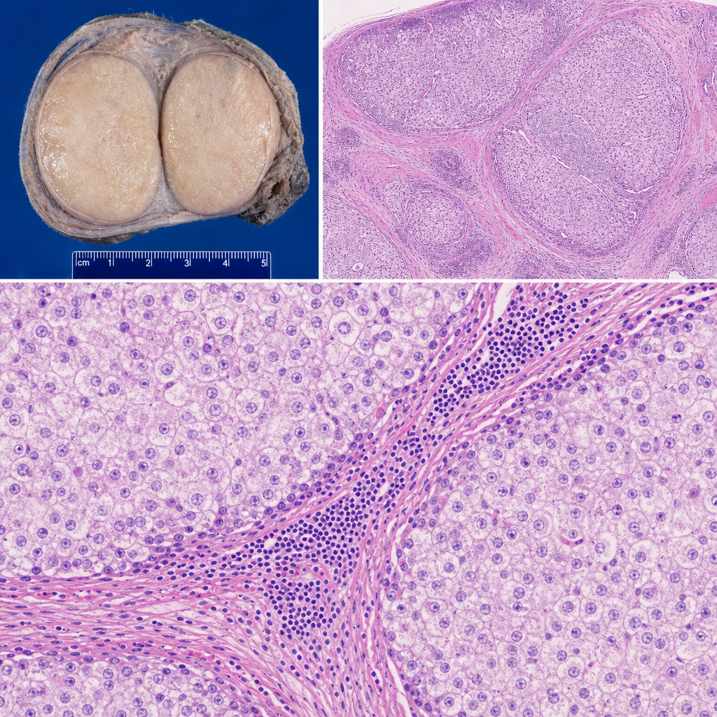
A 28-year-old man presents with a painless right testicular mass. Orchiectomy is performed. Gross examination reveals a 4 cm well-circumscribed, homogeneous, pale tan-gray mass without hemorrhage or necrosis. Histological sections show a monotonous population of large cells with clear, glycogen-rich cytoplasm and central round nuclei with prominent nucleoli, arranged in nests separated by fibrous septa infiltrated by mature lymphocytes. Which of the following best explains why the lymphocytic infiltrate is present within the fibrous septa of this tumor?
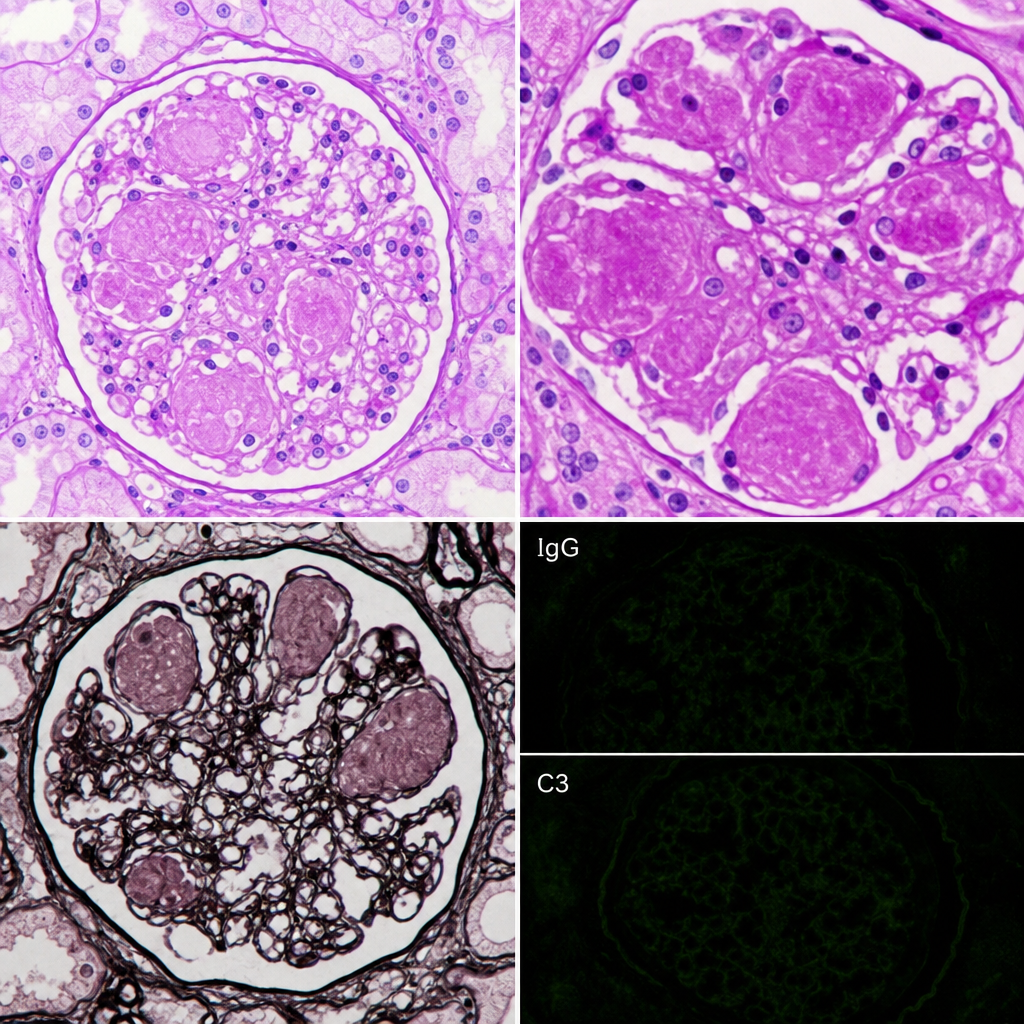
A 62-year-old man with poorly controlled type 2 diabetes mellitus for 18 years presents with proteinuria of 4.2 g/day and a serum creatinine of 2.8 mg/dL. Renal biopsy is performed. The biopsy shows PAS-positive, homogeneous, acellular nodular deposits in the mesangium of glomeruli, with associated capillary loop obliteration and diffuse glomerular basement membrane thickening. Immunofluorescence is negative for immune complex deposits and monoclonal light chain staining. Which of the following pathological processes best explains the formation of the nodular deposits seen in this biopsy?
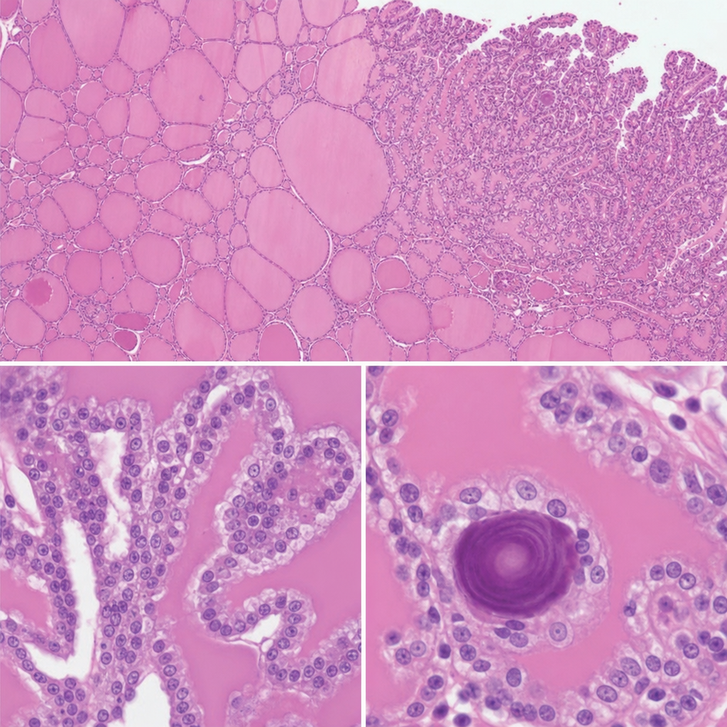
A 34-year-old woman undergoes excision of a neck mass. Histological examination of the resected specimen shows follicular structures of varying size filled with colloid, lined by cuboidal epithelium, with focal areas where small papillary projections bearing cells with ground-glass nuclei and nuclear grooves project into the follicular lumen. Scattered concentrically laminated calcified spherules are also identified within the papillary cores. Which of the following is the most likely origin and behavior of this neoplasm?
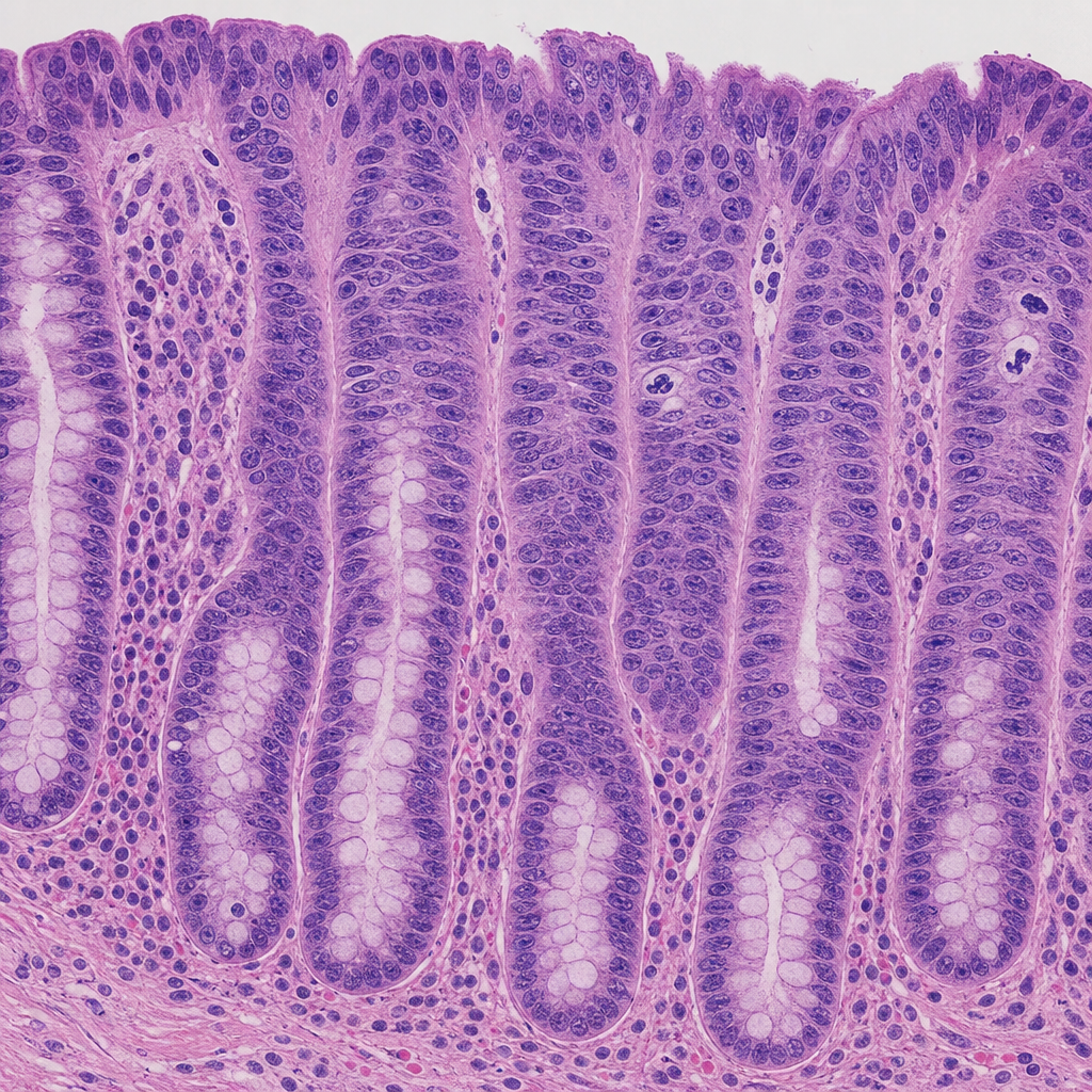
A 52-year-old man with a 25-year history of ulcerative colitis undergoes surveillance colonoscopy. A flat, velvety mucosal lesion is identified in the sigmoid colon and biopsied. The photomicrograph shows colonic crypts lined by cells with enlarged, hyperchromatic, stratified nuclei that extend to the luminal surface; the nuclei-to-cytoplasm ratio is markedly increased; goblet cells are absent; and mitotic figures including atypical forms are present in the upper third of the crypts. The basement membrane is intact with no stromal desmoplasia. Which of the following most accurately characterizes this lesion according to WHO classification principles?
A 35-year-old woman presents with headaches and seizures. MRI shows a well-circumscribed, calcified frontal lobe mass. Histology reveals oligodendroglioma with 1p/19q codeletion and IDH1 mutation. She undergoes gross total resection. Two years later, surveillance MRI shows a new enhancing nodule at the resection margin. Biopsy shows increased mitotic activity, microvascular proliferation, and retained 1p/19q codeletion but new CDKN2A/B homozygous deletion. What is the most critical factor in determining management strategy?
Practice by Chapter
Cerebrovascular diseases
Practice Questions
CNS trauma
Practice Questions
CNS infections
Practice Questions
Demyelinating diseases
Practice Questions
Neurodegenerative diseases
Practice Questions
Alzheimer's disease pathology
Practice Questions
Parkinson's disease pathology
Practice Questions
Amyotrophic lateral sclerosis
Practice Questions
Primary CNS tumors
Practice Questions
Metastatic CNS tumors
Practice Questions
Peripheral nerve disorders
Practice Questions
Skeletal muscle diseases
Practice Questions
Congenital CNS malformations
Practice Questions
Want unlimited practice?
Get full access to all questions, explanations, and performance tracking.
Scan to download app