Anatomy
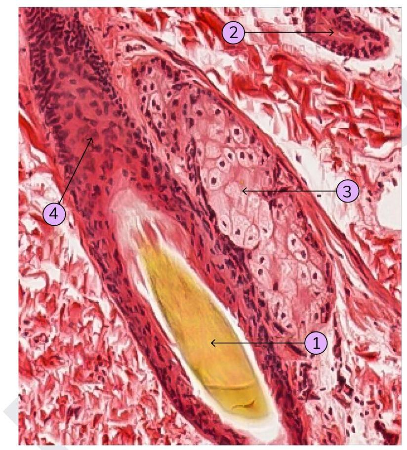
1 questionsIdentify which of the following structure is a sebaceous gland:
Dermatology
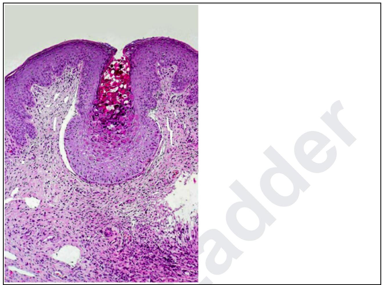
1 questionsA woman presents with lesions on the inner thighs and peri-anal region. They are nodular, 4-6 mm in size and appear pale. The histopathological image shows multiple intracytoplasmic inclusion bodies consistent with Henderson-Patterson bodies. The diagnosis is:
Pathology
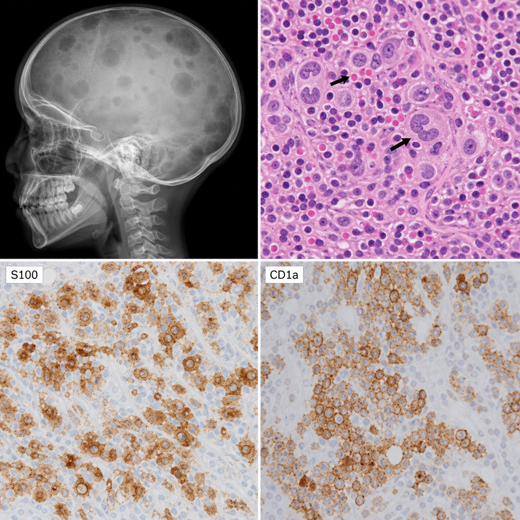
4 questionsA 7 year old child presents with fever, erythematous rash, and bone pain. $X$ - ray of the skull shows multiple lytic lesions. Skin biopsy shows prominent nuclear grooves with eosinophils as in the image below. Immunohistochemistry for S100, CD1a is also shown in the images below. What would be the diagnosis ?
Match the following cell types/patterns (Column A) with their associated malignancies (Column B): Column A (Cell types/patterns): a) Faggot cell b) Popcorn cell c) Starry sky pattern d) Cerebriform nuclei Column B (Associated malignancies): 1) Acute promyelocytic leukemia 2) Lymphocyte-predominant Hodgkin's lymphoma 3) Burkitt lymphoma 4) Sezary syndrome
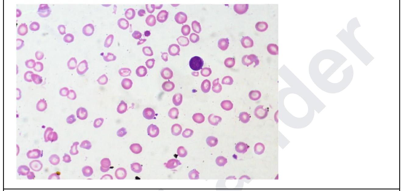
A 35 year old woman presents with fatigue. Investigations revealed the following: Hb, 5 g/dL; MCH, 24; low MCV; leukocytes, 11,000/ uL, and platelets, 5 lakhs. The peripheral smear is shown below. What is the diagnosis? Normal values: - Mean cell volume (MCV); 90 ± 8 fL - Mean cell Hb(MCH); 30 ± 3 pg
Iron in tissues is stained by:
Physiology
3 questionsWhich of the following is the primary tissue dependent on insulin for glucose uptake?
What is the normal insensible water loss?
A woman must vomit whenever she eats spicy food. Arrange the sequence of events during vomiting. 1. LES is open and UES is closed 2. Strong contractions in the stomach 3. Inspiration against a closed glottis 4. Relaxation of the pyloric sphincter 5. LES opens and UES opens 6. Reverse peristalsis in the small intestine LES: Lower esophageal sphincter UES: Upper esophageal sphincter
Radiology
1 questionsWhich of the following findings are seen in a high-resolution CT scan of fungal pneumonia? 1. Interlobular septations 2. Peripheral wedge-shaped consolidation 3. Pleural effusion 4. Cavitatory lesions with surrounding ground glass opacities