
Fertility and Infertility — MCQs

On this page
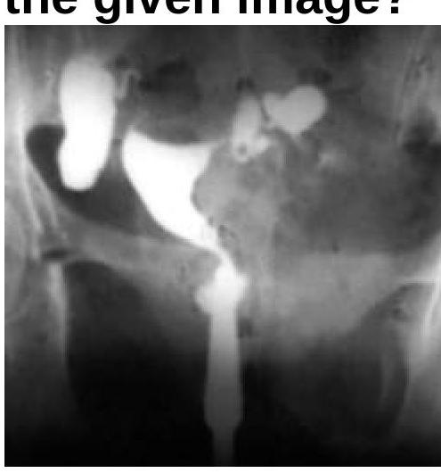
What will be the Hysterosalpingogram (HSG) finding?
Most common site for fertilization?
A 20 year old woman is evaluated for primary infertility. Hysterosalpingography was done and reveals an anomaly. What is the anomaly seen in the image?
What is meant by Superfecundation?
The window of implantation occurs at which of the following time periods after fertilization?
What is the most appropriate management for a 28-year-old hemodynamically stable patient with mild abdominal pain and an unruptured tubal ectopic pregnancy measuring 2.5 x 3 cm, with β-hCG level of 8500 mIU/mL, visible fetal cardiac activity, and who desires future fertility?
Which of the following is not considered a marker of ovarian reserve?
Which drug is commonly used in the medical management of ectopic pregnancy?
What does teratozoospermia refer to?
What is the most common presenting symptom of TB endometritis?
Fertility and Infertility Indian Medical PG Practice Questions and MCQs
Question 291: What will be the Hysterosalpingogram (HSG) finding?
- A. Hydrosalpinx (Correct Answer)
- B. Cornual block
- C. Normal findings
- D. Bicornuate uterus
Explanation: ***Hydrosalpinx*** - The image, likely a hysterosalpingogram (HSG), shows a **dilated and fluid-filled fallopian tube** with no spillage of contrast into the peritoneal cavity, which is characteristic of hydrosalpinx. - The **contrast media fills the tubal lumen** but is unable to egress, indicating distal tubal obstruction and fluid accumulation. *Cornual block* - A cornual block would present as **obstruction at the uterine ostium** of the fallopian tube, preventing contrast from entering the tubal lumen. - In this image, contrast has clearly entered and dilated the fallopian tube, ruling out a cornual block. *Normal findings* - Normal HSG findings would show **patent fallopian tubes** with free spill of contrast into the peritoneal cavity. - The visible **dilation** and **lack of spill** in the image are distinctly abnormal. *Bicornuate uterus* - A bicornuate uterus is a **congenital uterine anomaly** characterized by two separate uterine horns. - While the uterus appears somewhat irregular, the dominant feature is the dilated fallopian tube, which is not a hallmark of a bicornuate uterus.
Question 292: Most common site for fertilization?
- A. Isthmus
- B. Intramural
- C. Fimbriae
- D. Ampulla (Correct Answer)
Explanation: ***Ampulla*** - The **ampulla** of the **fallopian tube** is the widest and longest section, providing an ideal environment for the sperm and ovum to meet. - Fertilization most commonly occurs here, as it allows sufficient time for sperm capacitation and interaction with the egg. *Isthmus* - The **isthmus** is a narrow, thick-walled section of the fallopian tube, closer to the uterus. - While sperm may pass through here, it is not the primary site for fertilization. *Intramural* - The **intramural** (or interstitial) part is the segment of the fallopian tube that passes through the muscular wall of the uterus. - This narrowest part is not conducive to fertilization. *Fimbriae* - The **fimbriae** are finger-like projections at the end of the fallopian tube that capture the ovum after ovulation. - Their role is to direct the egg into the tube, not to be the site of fertilization.
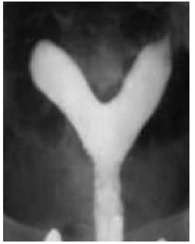
Question 293: A 20 year old woman is evaluated for primary infertility. Hysterosalpingography was done and reveals an anomaly. What is the anomaly seen in the image?
- A. Septate uterus (Correct Answer)
- B. Uterine didelphys
- C. Bicornuate uterus
- D. Unicornuate uterus
Explanation: ***Septate uterus*** - The image exhibits a **single uterine cavity** with a **septum** or indentation extending downwards, splitting the cavity into two distinct portions superiorly. - This configuration, particularly with an external contour that is typically **convex or flat**, is characteristic of a septate uterus, which is often associated with recurrent pregnancy loss and infertility. *Uterine didelphys* - This anomaly involves **two completely separate uteri**, each with its own cervix and often a separate vagina. - The image clearly shows a single main uterine body that then divides superiorly, not two entirely distinct uteri. *Bicornuate uterus* - A bicornuate uterus typically has two uterine horns that are **divergent externally**, creating a **deep indentation** on the external contour of the fundus. - While it also involves a divided uterine cavity, the external contour in the image appears more convex or flat, which is less consistent with a bicornuate uterus where the outer fundal contour is notably indented. *Unicornuate uterus* - This anomaly results from the **failure of one Müllerian duct to develop**, leading to a uterus that has only one horn and one fallopian tube. - The image presents a uterus with two distinct horns, ruling out a unicornuate uterus.
Question 294: What is meant by Superfecundation?
- A. Fertilization of ova and then its division
- B. Fertilization of two or more ova in one intercourse
- C. Fertilization of two or more ova in different intercourses in same menstrual cycle (Correct Answer)
- D. Fertilization of second ovum after first one is already implanted
Explanation: ***Fertilization of two or more ova in different intercourses in same menstrual cycle*** - **Superfecundation** occurs when two or more ova released during the same menstrual cycle are fertilized by sperm from **separate acts of coitus**. - This can lead to **dizygotic twins** or multiples conceived at different times within a short window, potentially from different biological fathers. *Fertilization of two or more ova in one intercourse* - This scenario describes the fertilization of multiple ova within a **single sexual encounter**, often leading to **dizygotic multiples** but not superfecundation. - Superfecundation specifically implies fertilization from **separate instances of intercourse**. *Fertilization of ova and then its division* - This describes the formation of **monozygotic (identical) twins**, where a single fertilized ovum (zygote) later splits into two or more embryos. - It is distinct from superfecundation, which involves fertilization of **multiple ova**. *Fertilization of second ovum after first one is already implanted* - This describes **superfetation**, a rare phenomenon where a new pregnancy (fertilization and conception) occurs **while already pregnant** from a previous cycle. - Superfecundation, conversely, involves **multiple conceptions within the same menstrual cycle**, not across different cycles.
Question 295: The window of implantation occurs at which of the following time periods after fertilization?
- A. 6-10 days (Correct Answer)
- B. 12 days
- C. 12 weeks
- D. 6 weeks
Explanation: ***6-10 days*** - The uterus is most receptive to implantation during the **"window of implantation,"** which occurs roughly **6 to 10 days post-fertilization**, coinciding with the mid-luteal phase. - During this period, the **endometrial lining** undergoes specific changes, guided by hormonal signals from **progesterone**, making it optimal for the blastocyst to attach. *12 days* - While implantation can still occur, the **peak receptivity window** is generally considered to be narrower, between 6 and 10 days. - By day 12, changes in the **endometrial environment** may start to reduce the likelihood of successful implantation. *12 weeks* - **12 weeks** refer to the end of the first trimester of pregnancy and is far too late for the initial implantation event. - Implantation must have occurred much earlier for a viable pregnancy at this stage. *6 weeks* - **6 weeks** refers to an established pregnancy, at which point implantation would have occurred several weeks prior. - The process of implantation is completed within the first two weeks post-fertilization.
Question 296: What is the most appropriate management for a 28-year-old hemodynamically stable patient with mild abdominal pain and an unruptured tubal ectopic pregnancy measuring 2.5 x 3 cm, with β-hCG level of 8500 mIU/mL, visible fetal cardiac activity, and who desires future fertility?
- A. Methotrexate therapy
- B. Laparoscopic salpingostomy (Correct Answer)
- C. Laparoscopic salpingectomy
- D. Expectant management
Explanation: ***Laparoscopic salpingostomy*** - This patient desires future fertility, making **salpingostomy** (tube-preserving surgery) the most appropriate management. - Salpingostomy involves making an incision in the fallopian tube, removing the ectopic pregnancy, and leaving the tube intact to preserve fertility potential. - While the presence of **fetal cardiac activity** and **β-hCG of 8500 mIU/mL** contraindicate medical management, they do not contraindicate conservative surgical management in a hemodynamically stable patient. - The patient meets criteria for conservative surgery: hemodynamically stable, unruptured ectopic, and desires future fertility. *Methotrexate therapy* - This patient has **absolute contraindications for methotrexate**: β-hCG level >5000 mIU/mL (here 8500) and presence of **fetal cardiac activity**. - Methotrexate is only suitable for hemodynamically stable patients with ectopic mass <3.5-4 cm, β-hCG <5000 mIU/mL, no fetal cardiac activity, and normal liver/renal function. - The high β-hCG and cardiac activity indicate a viable ectopic pregnancy that is unlikely to respond to medical management. *Laparoscopic salpingectomy* - Salpingectomy involves **complete removal of the affected fallopian tube**, which significantly reduces future fertility if this is the only functional tube or if the contralateral tube is damaged. - This option is preferred when: the tube is severely damaged, there is uncontrolled bleeding, recurrent ectopic in the same tube, or the patient does not desire future fertility. - Since this patient **specifically desires future fertility** and is hemodynamically stable with an unruptured ectopic, salpingostomy (tube preservation) is preferred over salpingectomy. *Expectant management* - Expectant management requires **very low or declining β-hCG levels** (typically <1000-1500 mIU/mL), absence of fetal cardiac activity, and very small ectopic size (<2 cm). - This patient has β-hCG of 8500 mIU/mL with **visible fetal cardiac activity**, indicating a viable growing ectopic pregnancy with high rupture risk. - These findings make expectant management unsafe and inappropriate.
Question 297: Which of the following is not considered a marker of ovarian reserve?
- A. Ovarian volume
- B. Inhibin B
- C. Anti-Müllerian Hormone (AMH)
- D. Inhibin A (Correct Answer)
Explanation: ***Inhibin A*** - **Inhibin A** levels primarily rise during the mid to late luteal phase and are involved in regulating FSH, but they are not a reliable or commonly used marker for **ovarian reserve**. - Its fluctuations are more indicative of the presence of a **corpus luteum** and short-term ovarian function rather than the total follicular pool. *Inhibin B* - **Inhibin B** is produced by granulosa cells of small antral follicles and is an important marker of **ovarian reserve**. - It inversely correlates with **FSH** levels in the early follicular phase, reflecting the number of developing follicles. *Ovarian volume* - **Ovarian volume**, particularly when measured by ultrasound, can be an indicator of **ovarian reserve**. - Smaller ovarian volume is generally associated with a reduced number of **antral follicles** and lower ovarian reserve. *Anti-Müllerian Hormone (AMH)* - **AMH** is a well-established and highly reliable marker of **ovarian reserve**, produced by the granulosa cells of preantral and small antral follicles. - Its levels correlate directly with the total number of remaining **primordial follicles** and are relatively stable throughout the menstrual cycle.
Question 298: Which drug is commonly used in the medical management of ectopic pregnancy?
- A. Leuprolide
- B. Methotrexate (Correct Answer)
- C. Mifepristone
- D. Carboprost
Explanation: ***Correct: Methotrexate*** - **Methotrexate** is a **folic acid antagonist** that inhibits DNA synthesis and cell proliferation, making it effective in terminating early ectopic pregnancies by targeting rapidly dividing trophoblastic cells. - It is typically considered for **hemodynamically stable** patients with unruptured ectopic pregnancies, a beta-hCG level below a certain threshold (e.g., <5,000 mIU/mL), and no cardiac activity in the ectopic mass. - This is the **gold standard** for medical management of ectopic pregnancy meeting specific criteria. *Incorrect: Mifepristone* - **Mifepristone** is an **antiprogestin** primarily used for medical abortion of intrauterine pregnancies, causing detachment of the gestational sac and cervical ripening. - While it can be used in combination with misoprostol for medical abortion, it is **not the primary drug** for managing ectopic pregnancies. *Incorrect: Leuprolide* - **Leuprolide** is a **GnRH agonist** mainly used for conditions like endometriosis, uterine fibroids, and prostate cancer by suppressing ovarian or testicular hormone production. - It is **not used** in the direct medical management of ectopic pregnancy. *Incorrect: Carboprost* - **Carboprost** is a **prostaglandin F2-alpha analog** primarily used to treat **postpartum hemorrhage** by inducing strong uterine contractions. - It is **not indicated** for the treatment of ectopic pregnancy.
Question 299: What does teratozoospermia refer to?
- A. Low sperm count
- B. Sperm with abnormal motility
- C. Absence of sperm in semen
- D. Morphologically defective sperm (Correct Answer)
Explanation: ***Morphologically defective sperm*** - **Teratozoospermia** specifically refers to the presence of an unusually high percentage of **abnormally shaped sperm** in an ejaculate. - These malformations can affect the **head, midpiece, or tail** of the sperm, potentially impairing its ability to fertilize an egg. *Low sperm count* - This condition is known as **oligozoospermia**, which refers to a sperm concentration below the normal range. - While low sperm count can affect fertility, it is distinct from issues with sperm morphology. *Sperm with abnormal motility* - This condition is called **asthenozoospermia**, characterized by reduced or absent sperm movement. - Poor motility impacts the sperm's ability to reach and penetrate the egg, but it is not directly related to sperm shape. *Absence of sperm in semen* - The complete absence of sperm in the ejaculate is known as **azoospermia**. - This is a severe form of male infertility, different from having sperm with structural defects.
Question 300: What is the most common presenting symptom of TB endometritis?
- A. Amenorrhoea
- B. Vaginal discharge
- C. Abdominal pain
- D. Infertility (Correct Answer)
Explanation: ***Infertility*** - **Infertility** is the most common presenting symptom of **tuberculosis (TB) endometritis**, particularly secondary infertility. - The infection leads to inflammation and scarring of the endometrium and fallopian tubes, impairing implantation and ovum transport. *Abdominal pain* - While **abdominal pain** can occur in TB endometritis, it is typically a less frequent or prominent presenting symptom compared to infertility. - Pain often arises from pelvic inflammation or adhesions but is not the cardinal complaint that prompts diagnosis. *Amenorrhoea* - **Amenorrhea** (absence of menstruation) can be a symptom, especially in advanced cases where there is significant destruction of the endometrium. - It is, however, less common than infertility as the initial presenting symptom. *Vaginal discharge* - **Vaginal discharge** is an uncommon symptom of TB endometritis. - When present, it is often non-specific and not characteristic enough to suggest TB as the underlying cause.
Practice by Chapter
Reproductive Physiology
Practice Questions
Evaluation of the Infertile Couple
Practice Questions
Male Factor Infertility
Practice Questions
Female Factor Infertility
Practice Questions
Ovulatory Disorders
Practice Questions
Tubal and Peritoneal Factors
Practice Questions
Uterine Factors
Practice Questions
Unexplained Infertility
Practice Questions
Assisted Reproductive Technologies
Practice Questions
Psychological Aspects of Infertility
Practice Questions
Want unlimited practice?
Get full access to all questions, explanations, and performance tracking.
Start For Free