
Trauma — MCQs

On this page
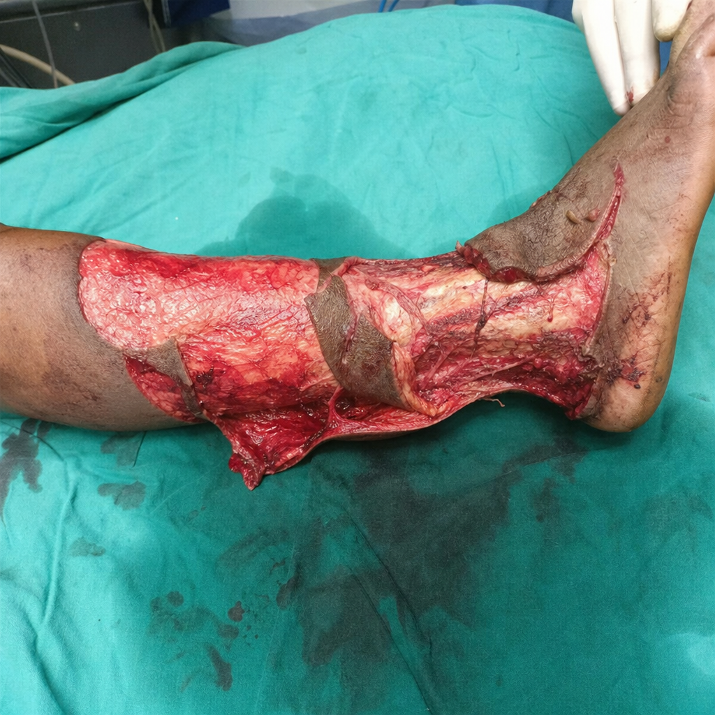
To which category will you classify this wound?
Bullet wounds near major blood vessels should be explored only if -
A 22-year-old woman is found in a comatose condition, having lain for an unknown length of time on the tile floor of the courtyard. She is found in possession of cocaine. The patient is transported to the hospital while EMT personnel receive instructions for treatment of drug overdose. During the physical examination the patient's gluteal region shows signs of ischemia. After regaining consciousness, she exhibits paralysis of knee flexion and dorsal and plantar flexion and sensory loss in the limb. What is the most likely diagnosis?
Fluid of choice for resuscitation of a burn patient:
A patient presents to the ER after an RTA. What is the best way to differentiate cardiac tamponade from tension pneumothorax?
When resuscitating a patient in shock which of the following is not an adequate parameter to predict end point of resuscitation?
A man comes to the emergency department with stab injury to left flank. He has stable vitals. What would be the next step in management?
What is the % Body surface area involved in burns of the perineum?
Which of the following is false regarding first degree burns
Which is not an indication for thoracotomy?
Practice by Chapter
Initial Assessment of Trauma Patient
Practice Questions
Advanced Trauma Life Support (ATLS) Principles
Practice Questions
Chest Trauma
Practice Questions
Abdominal Trauma
Practice Questions
Head Trauma
Practice Questions
Spinal Trauma
Practice Questions
Extremity Trauma
Practice Questions
Vascular Trauma
Practice Questions
Genitourinary Trauma
Practice Questions
Burns Management
Practice Questions
Mass Casualty Management
Practice Questions
Damage Control Surgery
Practice Questions
Want unlimited practice?
Get full access to all questions, explanations, and performance tracking.
Scan to download app