
Maternal-Fetal Medicine — MCQs

On this page
The major contributor to amniotic fluid after 20 weeks of gestation is:
Chromosome number of partial hydatidiform mole is-
Which of the following statement is correct about acute fatty liver of pregnancy?
What maternal condition is commonly associated with congenital heart defects in the fetus?
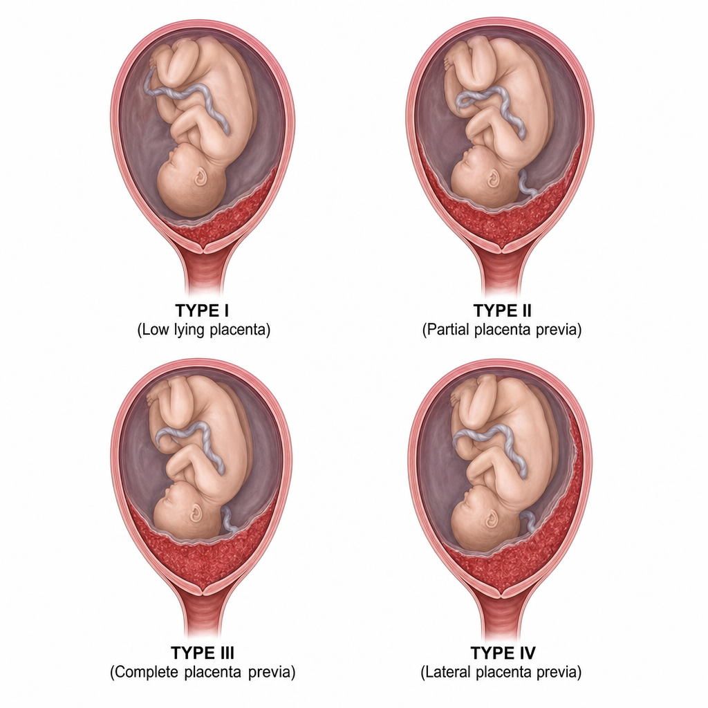
⚠️ [IMAGE MISSING] Identify the type of placenta praevia based on the provided image.
A 22-year-old primigravida visits ANC OPD with 20 weeks POG. On examination uterine height reveals a 16-week size. USG shows reduced liquor. What will be the diagnosis?
A female patient collapses soon after delivery. There is profuse bleeding and features of disseminated intravascular coagulation. Which of the following is the most likely etiology?
A primigravida at 22 weeks of gestation presents with profuse vaginal bleeding. Her blood pressure and glucose levels are normal. At which of the following sites can placental implantation lead to this condition?
In a case of DCDA twins at 38 weeks, with the first twin in breech presentation, and the mother having a blood pressure of 140/96 and 1+ proteinuria, what is the preferred management?
A patient with recurrent abortion is diagnosed to have antiphospholipid syndrome. What will be the treatment?
Practice by Chapter
Fetal Assessment Techniques
Practice Questions
Hypertensive Disorders in Pregnancy
Practice Questions
Intrauterine Growth Restriction
Practice Questions
Multiple Gestation
Practice Questions
Rh Isoimmunization and Other Blood Group Incompatibilities
Practice Questions
Intrauterine Fetal Therapy
Practice Questions
Prenatal Diagnosis and Genetic Counseling
Practice Questions
Placental Abnormalities
Practice Questions
Preterm Labor and Delivery
Practice Questions
Management of Medical Disorders in Pregnancy
Practice Questions
Want unlimited practice?
Get full access to all questions, explanations, and performance tracking.
Scan to download app