
Systemic Anatomy — MCQs

On this page
Which of the following structures of joints is not innervated?
Cremasteric artery is a branch of?
Which of the following is a retroperitoneal structure?
Inferior epigastric vein drains into?
Appendices epiploicae are a feature of?
Valve of Heister is seen in
Inferior rectal artery is a branch of?
Which of the following is not a branch of the lumbar plexus?
Superficial epigastric artery is a branch of?
Which of the following structures is not a boundary of Calot's triangle shown in the given image?
Systemic Anatomy Indian Medical PG Practice Questions and MCQs
Question 231: Which of the following structures of joints is not innervated?
- A. Synovium
- B. Ligaments
- C. Capsule
- D. Articular cartilage (Correct Answer)
Explanation: ***Articular cartilage*** - **Articular cartilage** is primarily composed of **chondrocytes** embedded in an extracellular matrix, lacking **nerves** and **blood vessels** [1]. - Its **aneural** nature explains why damage to articular cartilage often causes no direct pain until underlying structures are affected [1]. *Synovium* - The **synovial membrane** is richly innervated with **nociceptors** and **mechanoreceptors**, contributing to pain perception and proprioception within joints. - Inflammation of the synovium (**synovitis**) is a common cause of joint pain. *Capsule* - The **fibrous capsule** surrounding a joint is densely innervated by **sensory nerve endings**, including **nociceptors** and **mechanoreceptors**. - Stretching or damage to the joint capsule can result in significant pain. *Ligaments* - **Ligaments** are **well-innervated** with sensory nerve endings, particularly **proprioceptors** and **nociceptors**. - This innervation allows ligaments to provide feedback on joint position and contribute to pain sensation upon injury.
Question 232: Cremasteric artery is a branch of?
- A. Internal pudendal artery
- B. External pudendal artery
- C. Inferior epigastric artery (Correct Answer)
- D. Superior epigastric artery
Explanation: ***Inferior epigastric artery*** - The **cremasteric artery** (also known as the external spermatic artery) is a branch of the **inferior epigastric artery**. - It supplies the **cremaster muscle** and other structures within the spermatic cord. *Internal pudendal artery* - The **internal pudendal artery** primarily supplies the perineum and external genitalia. - It does not typically give rise to the cremasteric artery. *External pudendal artery* - The **external pudendal artery** typically supplies the skin of the scrotum/labia majora and the perineum [2]. - It is not the origin of the cremasteric artery. *Superior epigastric artery* - The **superior epigastric artery** is a terminal branch of the internal thoracic artery and supplies the upper part of the anterior abdominal wall [1]. - It is anatomically distant and unrelated to the origin of the cremasteric artery.
Question 233: Which of the following is a retroperitoneal structure?
- A. Ileum
- B. Jejunum
- C. Ureter (Correct Answer)
- D. Appendix
Explanation: ***Ureter*** - The **ureters** are tubes that carry urine from the kidneys to the bladder and are located **posterior to the peritoneum**, making them retroperitoneal structures [1]. - This anatomical position means they are covered anteriorly by the peritoneum but not suspended by a mesentery [2]. *Ileum* - The **ileum** is part of the small intestine and is an **intraperitoneal organ**, meaning it is suspended within the abdominal cavity by the mesentery. - Its peritoneal covering allows for significant mobility within the abdomen. *Jejunum* - Like the ileum, the **jejunum** is also an **intraperitoneal organ**, suspended by the mesentery and allowing for free movement. - It is located within the greater sac of the peritoneal cavity. *Appendix* - The **appendix** is typically an **intraperitoneal structure**, suspended by its own mesentery, the mesoappendix. - While it can be located in a retrocecal position (behind the cecum), it remains primarily an intraperitoneal organ due to its peritoneal covering.
Question 234: Inferior epigastric vein drains into?
- A. Femoral vein
- B. External iliac vein (Correct Answer)
- C. Internal iliac vein
- D. Internal pudendal vein
Explanation: ***External iliac vein*** - The **inferior epigastric vein** runs superiorly from the **inguinal ligament** and is a direct tributary of the external iliac vein, which then continues as the common iliac vein. [2] - This anatomical connection is crucial in understanding the venous drainage of the **anterior abdominal wall** inferior to the umbilicus. [2] *Femoral vein* - The **femoral vein** is a continuation of the popliteal vein and drains the lower limb, eventually becoming the external iliac vein above the inguinal ligament. [1] - The inferior epigastric vein does **not directly drain** into the femoral vein. *Internal iliac vein* - The **internal iliac vein** primarily drains structures within the **pelvis** and the **gluteal region**. - It does not receive direct drainage from the inferior epigastric vein, which is associated with the anterior abdominal wall. [2] *Internal pudendal vein* - The **internal pudendal vein** drains structures of the **perineum** and parts of the external genitalia. - It is a tributary of the internal iliac vein and plays no direct role in draining the inferior epigastric region.
Question 235: Appendices epiploicae are a feature of?
- A. Duodenum
- B. Stomach
- C. Colon (Correct Answer)
- D. Jejunum
Explanation: ***Colon*** - **Appendices epiploicae** are small, fat-filled pouches of peritoneum attached to the outer surface of the **colon**, distinguishing it from other parts of the gastrointestinal tract. - They are most numerous and prominent on the **transverse** and **sigmoid colon** [1]. *Duodenum* - The **duodenum** is the first part of the small intestine and lacks appendices epiploicae. - Its distinguishing features include **Brunner's glands** in the submucosa. *Stomach* - The **stomach** is characterized by rugae (folds of the mucosa) and multiple muscle layers, but it does not have appendices epiploicae. - Its primary function is mechanical and chemical digestion of food. *Jejunum* - The **jejunum**, part of the small intestine, is characterized by prominent **plica circulares (circular folds)** and long villi, but it does not possess appendices epiploicae. - It is mainly involved in nutrient absorption [2].
Question 236: Valve of Heister is seen in
- A. Cystic duct (Correct Answer)
- B. Common bile duct
- C. Common hepatic duct
- D. Pancreatic duct
Explanation: ***Cystic duct*** - The **spiral valve of Heister** (or Valves of Heister) are a series of crescentic folds of mucous membrane found within the cystic duct [1]. - These valves are thought to help prevent the collapse or over-distension of the cystic duct, ensuring a continuous flow of bile to and from the gallbladder. *Common bile duct* - The common bile duct is formed by the union of the **cystic duct** and the **common hepatic duct**. - It does not contain the Valve of Heister; its primary function is to transport bile to the duodenum. *Common hepatic duct* - The common hepatic duct is formed by the union of the **right and left hepatic ducts** from the liver. - It also does not contain the Valve of Heister; its role is to drain bile from the liver. *Pancreatic duct* - The pancreatic duct (or **Duct of Wirsung**) carries digestive enzymes from the pancreas to the duodenum. - It is anatomically distinct from the biliary system and does not contain the Valve of Heister.
Question 237: Inferior rectal artery is a branch of?
- A. Inferior mesenteric artery
- B. Superior mesenteric artery
- C. Coeliac trunk
- D. Internal pudendal artery (Correct Answer)
Explanation: ***Internal pudendal artery*** - The **inferior rectal artery** is a direct branch of the internal pudendal artery which supplies the anal canal [1]. - The **internal pudendal artery** is a branch of the internal iliac artery, supplying structures in the perineum [1]. *Inferior mesenteric artery* - The inferior mesenteric artery supplies the **distal transverse colon**, descending colon, sigmoid colon, and superior part of the rectum (via the superior rectal artery) [1]. - It does not directly supply the lower anal canal, which is the domain of the inferior rectal artery. *Superior mesenteric artery* - The superior mesenteric artery supplies the **small intestine** and the **proximal large intestine** up to the transverse colon [1]. - It supplies the midgut derivatives and is not involved in supplying the rectum or anal canal directly. *Coeliac trunk* - The coeliac trunk is the main arterial supply to the **foregut** organs, including the stomach, liver, spleen, and pancreas [1]. - It does not supply any part of the rectum or anal canal.
Question 238: Which of the following is not a branch of the lumbar plexus?
- A. Ilioinguinal nerve
- B. Genitofemoral nerve
- C. Iliohypogastric nerve
- D. Subcostal nerve (Correct Answer)
Explanation: ***Subcostal nerve*** - The **subcostal nerve** is the ventral ramus of the **T12 spinal nerve** and is therefore part of the **thoracic spinal nerves**, not the lumbar plexus. - It runs inferior to the 12th rib, innervates the **external oblique muscle**, and contributes to the innervation of the **rectus abdominis** and **pyramidalis muscles**. *Iliohypogastric nerve* - This nerve is a branch of the **lumbar plexus** (L1) and innervates the **internal oblique** and **transversus abdominis muscles** [1]. - It also provides cutaneous innervation to the **suprapubic region** and a small part of the buttock. *Ilioinguinal nerve* - The ilioinguinal nerve is also a branch of the **lumbar plexus** (L1) and runs parallel to the iliohypogastric nerve [1]. - It provides cutaneous innervation to the **upper medial thigh**, base of the penis/mons pubis, and the anterior part of the scrotum/labium majus. *Genitofemoral nerve* - Originating from the **lumbar plexus** (L1, L2), the genitofemoral nerve divides into a **genital branch** and a **femoral branch**. - The genital branch innervates the **cremaster muscle** in males and the **labia majora** in females, while the femoral branch supplies cutaneous innervation to the **anterior thigh**.
Question 239: Superficial epigastric artery is a branch of?
- A. Internal pudendal artery
- B. External pudendal artery
- C. Internal iliac artery
- D. Femoral artery (Correct Answer)
Explanation: ***Femoral artery*** - The **superficial epigastric artery** is one of the initial branches that arises directly from the **femoral artery** in the femoral triangle, just below the inguinal ligament. - It supplies the **skin** and **superficial fascia** over the lower part of the anterior abdominal wall and the superficial inguinal lymph nodes [1]. *Internal pudendal artery* - The **internal pudendal artery** is a branch of the **internal iliac artery** and primarily supplies structures in the perineum and genitalia. - It does not supply the anterior abdominal wall. *External pudendal artery* - The **external pudendal artery** is also a branch of the **femoral artery**, but it supplies the skin of the external genitalia (scrotum/labia majora) and adjacent thigh. - It does not supply the superficial epigastric region. *Internal iliac artery* - The **internal iliac artery** primarily supplies the pelvic organs, gluteal region, and medial thigh. - While it has many branches, the superficial epigastric artery is not one of them.
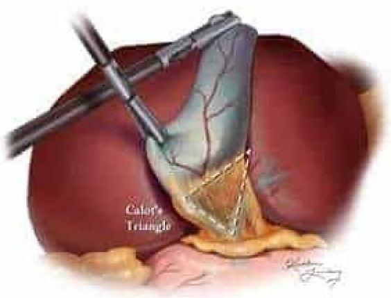
Question 240: Which of the following structures is not a boundary of Calot's triangle shown in the given image?
- A. Common hepatic duct
- B. Cystic duct
- C. Inferior surface of the liver
- D. Gallbladder (Correct Answer)
Explanation: ***Gallbladder*** - The image depicts **Calot's triangle**, which is an important anatomical landmark in gallbladder surgery. The gallbladder itself is located within this region, but it is not one of the defined boundaries of the triangle. - While central to the anatomy shown, the **gallbladder** is surrounded by the structures that form the triangle's boundaries rather than bounding it itself. *Common hepatic duct* - The **common hepatic duct** forms the medial boundary of Calot's triangle. - This duct is formed by the union of the right and left hepatic ducts and carries bile from the liver. *Cystic duct* - The **cystic duct** forms the lateral (or inferior) boundary of Calot's triangle. - This duct connects the gallbladder to the common hepatic duct. *Inferior surface of the liver* - The **inferior surface of the liver** forms the superior boundary of Calot's triangle. - Specifically, this refers to the edge of the right lobe of the liver at the base of the gallbladder fossa.
Practice by Chapter
Skeletal System
Practice Questions
Articular System
Practice Questions
Muscular System
Practice Questions
Cardiovascular System
Practice Questions
Lymphatic System
Practice Questions
Nervous System
Practice Questions
Respiratory System
Practice Questions
Digestive System
Practice Questions
Urinary System
Practice Questions
Reproductive System
Practice Questions
Endocrine System
Practice Questions
Integumentary System
Practice Questions
Want unlimited practice?
Get full access to all questions, explanations, and performance tracking.
Start For Free