
Microscopic Anatomy — MCQs

On this page
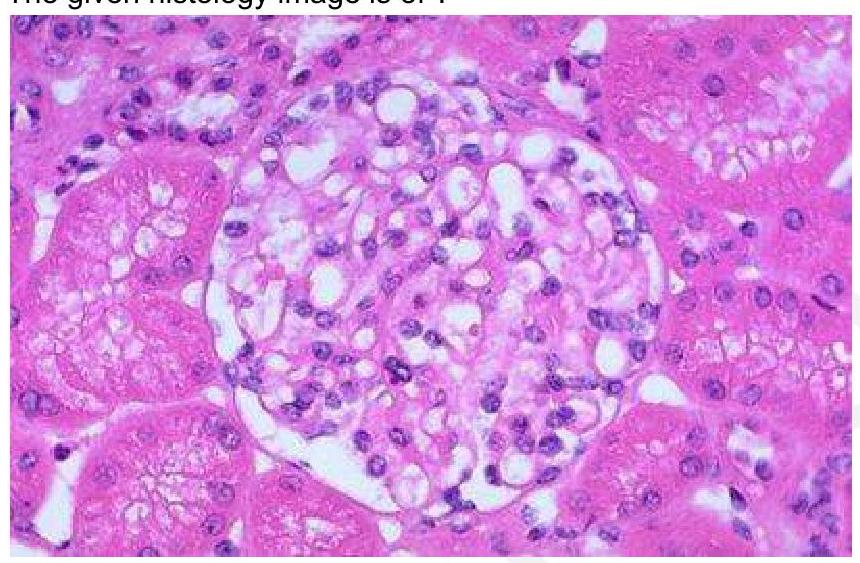
The given histology image is of which structure?
Cells which surround the oocyte in graafian follicle are called ?
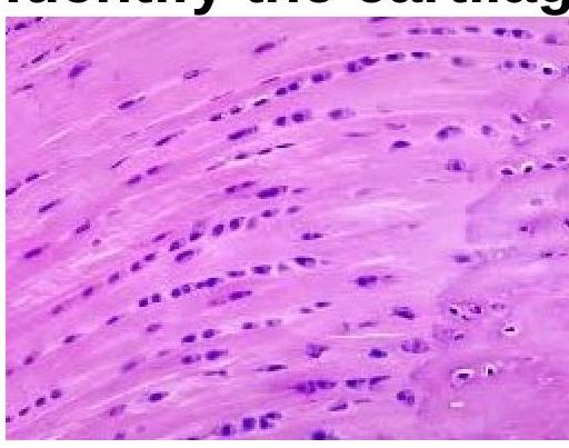
Identify the type of cartilage shown in the image.
Which type of collagen is the most abundant in the human skin?
Which of the following accurately describes the anatomical structure of the hard palate?
Ceruminous glands present in the ear are:
Which area in the spleen is considered *primarily* thymus-dependent?
What is the type of epithelium of the adenoid?
Intercalated disc is present in:
In the context of blood pressure regulation, where are baroreceptors primarily located?
Microscopic Anatomy Indian Medical PG Practice Questions and MCQs
Question 211: The given histology image is of which structure?
- A. Pancreatic islet cells
- B. Hassall's corpuscles
- C. Glomerulus (Correct Answer)
- D. Leydig cells in the testis
Explanation: ***Glomerulus*** - The glomerulus is characterized by a **tuft of capillaries** surrounded by Bowman's capsule, responsible for filtration in the kidney [1]. - Histological examination typically shows a **dense network of capillaries** and **mesangial cells**, which are distinct features of the glomeruli [1]. *Hassall's corpuscles* - Found in the **thymus**, they are round structures composed of epithelial cells, crucial in T-cell maturation. - Histologically, they present as concentric layers of **epithelial cells** and are not found in the kidney. *Leydig cells of testis* - Located in the **interstitial tissue** of the testes, these cells produce testosterone and are typically larger than glomerular cells. - They are characterized by their **eosinophilic cytoplasm** and round nuclei, differing markedly from the structures found in the glomerulus. *Pancreatic islet cells* - Islet cells are involved in **hormone production**, predominantly insulin and glucagon, and are located in the pancreas. - Histologically, they appear as small clusters dispersed among **exocrine pancreas**, which is different from the highly organized structure of the glomerulus. **References:** [1] Cross SS. Underwood's Pathology: A Clinical Approach. 6th ed. Common Clinical Problems From Diseases Of The Urinary And Male Genital Tracts, pp. 522-523.
Question 212: Cells which surround the oocyte in graafian follicle are called ?
- A. Discus proligerus
- B. Luteal cells
- C. Villus cells
- D. Cumulus oophorus (Correct Answer)
Explanation: ***Cumulus oophorus*** - The **cumulus oophorus** is a mound of **granulosa cells** that surrounds the oocyte in the Graafian follicle [2]. - These cells are crucial for the development, maturation, and eventual ovulation of the oocyte. *Discus proligerus* - **Discus proligerus** is an older, less commonly used term for the **cumulus oophorus**. - While it refers to the same structure, **cumulus oophorus** is the more current and preferred terminology. *Luteal cells* - **Luteal cells** are formed from the granulosa and theca cells of the ruptured follicle after ovulation, forming the **corpus luteum** [1]. - They are involved in hormone production (progesterone) after the oocyte has been released, not directly surrounding the oocyte within the intact Graafian follicle [1]. *Villus cells* - **Villus cells** are primarily found in the **placenta** and **intestines**, where they are involved in nutrient exchange and absorption. - They have no role in the structure or function of the ovarian follicle or the oocyte within it.
Question 213: Identify the type of cartilage shown in the image.
- A. Hyaline cartilage
- B. Elastic cartilage
- C. Dense connective tissue
- D. Fibrocartilage (Correct Answer)
Explanation: ***Fibrocartilage*** - The image displays **chondrocytes** arranged in rows between bundles of **collagen fibers**, characteristic of fibrocartilage. This structure provides high tensile strength and shock absorption. - Unlike other cartilage types, fibrocartilage lacks a distinct perichondrium and is found in structures requiring extreme strength, such as intervertebral discs and menisci. *Hyaline cartilage* - Hyaline cartilage has an amorphous, **glassy matrix** with chondrocytes typically arranged in small groups (isogenous groups) within lacunae, without prominent collagen bundles. - It is found in articular surfaces, tracheal rings, and the costal cartilages, providing smooth, low-friction surfaces. *Elastic cartilage* - Elastic cartilage is characterized by a high concentration of **elastic fibers** within its matrix, giving it flexibility and resilience. - Chondrocytes are typically scattered and the matrix stains darker due to the elastic fibers, which are not apparent in the image. *Dense connective tissue* - While dense connective tissue contains abundant **collagen fibers**, it lacks chondrocytes in lacunae, instead containing fibroblasts. - The presence of chondrocytes in the image confirms this is cartilage, not dense connective tissue.
Question 214: Which type of collagen is the most abundant in the human skin?
- A. Type II collagen
- B. Type I collagen (Correct Answer)
- C. Type III collagen
- D. Type IV collagen
Explanation: ***Type I collagen*** - **Type I collagen** is the **most abundant type** of collagen in the human body, accounting for about 90% of the collagen, and is particularly prevalent in the **dermis** of the skin, where it provides tensile strength and elasticity [1]. - It forms **thick, striated fibrils** and is critical for the structural integrity of skin, bones, tendons, and ligaments [1]. *Type II collagen* - **Type II collagen** is primarily found in **cartilage**, especially **hyaline and elastic cartilage**, where it provides resistance to pressure. - It is not a major component of the skin's dermal structure. *Type III collagen* - **Type III collagen** is found alongside Type I collagen in the skin but is prominent in **reticular fibers** and plays a role in the elasticity and suppleness of the skin, particularly in rapidly growing tissues and blood vessels. - While present in the skin, it is less abundant than Type I collagen and its primary role is in supporting organ structure, not tensile strength. *Type IV collagen* - **Type IV collagen** is a major component of **basement membranes**, forming a mesh-like network that provides structural support and acts as a filter in tissues. - It is not found in the interstitial connective tissue of the dermis but rather underlies the epidermis, forming part of the dermoepidermal junction.
Question 215: Which of the following accurately describes the anatomical structure of the hard palate?
- A. Non-keratinised mucosa with a submucosal layer and minor salivary glands.
- B. Non-keratinised mucosa without a submucosal layer and minor salivary glands.
- C. Keratinised mucosa without a submucosal layer and minor salivary glands.
- D. Keratinised mucosa with a submucosal layer and minor salivary glands. (Correct Answer)
Explanation: ***Keratinised mucosa with a submucosal layer and minor salivary glands.*** - The hard palate is covered by **keratinised stratified squamous epithelium** to withstand the forces of chewing and mastication. - It contains a **submucosal layer** in its posterolateral region (near the molar teeth) and anteriorly (near the incisor teeth) where **minor salivary glands** (palatine glands) are present. *Non-keratinised mucosa with a submucosal layer and minor salivary glands.* - The hard palate is primarily covered by **keratinised epithelium**, not non-keratinised, due to its role in food processing. - While it does have a submucosal layer and minor salivary glands in specific regions, the mucosal type is incorrect. *Non-keratinised mucosa without a submucosal layer and minor salivary glands.* - This option incorrectly states that the hard palate has **non-keratinised mucosa** and no submucosal layer, which is false for several regions. - The presence of minor salivary glands is also incorrect for this description. *Keratinised mucosa without a submucosal layer and minor salivary glands.* - While the hard palate is indeed covered by **keratinised mucosa**, it is incorrect to state that it entirely lacks a submucosal layer or minor salivary glands. - These structures are present in the posterolateral and anterior regions of the hard palate.
Question 216: Ceruminous glands present in the ear are:
- A. Modified eccrine glands
- B. Modified apocrine glands (Correct Answer)
- C. Mucous gland
- D. Modified holocrine glands
Explanation: ***Modified apocrine glands*** - **Ceruminous glands** in the ear canal are specialized **apocrine glands** responsible for producing cerumen (earwax). - Like other apocrine glands, they secrete their product via **apical budding** of the cell, releasing fragments of the cell along with the secretion [1]. *Modified eccrine glands* - **Eccrine sweat glands** are distributed throughout the skin and produce a watery sweat for thermoregulation [1]. - They secrete their product directly onto the skin surface via **exocytosis**, without loss of cellular material. *Mucous gland* - **Mucous glands** (e.g., salivary glands, respiratory tract glands) produce **mucus**, a viscous secretion primarily for lubrication and protection. - Their secretions are rich in **mucin glycoproteins**, which is distinct from the lipid-rich cerumen. *Modified holocrine glands* - **Holocrine glands** (e.g., sebaceous glands) release their entire cell contents, including lipids and cellular debris, upon cell lysis. - While sebaceous glands contribute to earwax, ceruminous glands themselves operate via an **apocrine mechanism**, not holocrine [1].
Question 217: Which area in the spleen is considered *primarily* thymus-dependent?
- A. Mantle layer
- B. Malpighian corpuscle
- C. Periarteriolar lymphoid sheath (PALS) (Correct Answer)
- D. None of the options
Explanation: ***Periarteriolar lymphoid sheath (PALS)*** - PALS is the **T-cell zone** of splenic white pulp, containing predominantly **T-lymphocytes** arranged around central arterioles. [2] - This area is **thymus-dependent** as it houses mature T cells that have undergone thymic selection and education, making it the primary thymus-dependent area of the spleen. [1] *Mantle layer* - The mantle layer consists of **naïve B lymphocytes** that surround the germinal center within splenic follicles. - This area is **thymus-independent** and primarily involved in **B-cell responses** to antigens. *Malpighian corpuscle* - Also known as **splenic follicles** or white pulp, this area primarily functions as **B-cell aggregation zones**. - While containing both B and T cell areas, it's predominantly **thymus-independent** with its main role being B-cell activation and antibody production. *None of the options* - This option is incorrect because PALS clearly represents the primary **thymus-dependent area** in the spleen. - The spleen definitively contains thymus-dependent zones where **T-cell activation** and proliferation occur, specifically the PALS. [1]
Question 218: What is the type of epithelium of the adenoid?
- A. Pseudostratified ciliated columnar epithelium (Correct Answer)
- B. Non-keratinized squamous epithelium
- C. Cuboidal epithelium
- D. Columnar epithelium with goblet cells
Explanation: ***Pseudostratified ciliated columnar epithelium*** - The adenoid (pharyngeal tonsil) is located in the nasopharynx, which is part of the upper respiratory tract and is lined with **pseudostratified ciliated columnar epithelium** with goblet cells [1]. - This type of epithelium is also known as **respiratory epithelium**, and its cilia and mucus-producing goblet cells help to trap and clear inhaled particles and pathogens [1]. *Non-keratinized squamous epithelium* - This type of epithelium is found in areas subject to friction and abrasion, such as the oral cavity, oro- and laryngopharynx, and esophagus, not typically in the nasopharynx. - It provides protection but lacks the ciliated and mucus-producing cells necessary for clearing respiratory passages. *Cuboidal epithelium* - This epithelium is typically found in glands and ducts, secretory and absorptive surfaces, such as renal tubules and thyroid follicles. - It does not have the specialized functions (cilia, mucus production) required for the respiratory system's protective lining. *Columnar epithelium with goblet cells* - While the adenoid epithelium does contain **goblet cells** for mucus production, specifically stating "columnar epithelium" is not as precise as "pseudostratified ciliated columnar epithelium." - The key feature of being **pseudostratified** and **ciliated** is crucial for its function in the nasopharynx [1].
Question 219: Intercalated disc is present in:
- A. Cardiac muscle (Correct Answer)
- B. Smooth muscle
- C. Skeletal muscle
- D. All of the options
Explanation: ***Cardiac muscle*** - **Intercalated discs** are unique structures found only in cardiac muscle, forming specialized cell-cell junctions [1]. - These discs contain **gap junctions** for electrical coupling and **desmosomes** for strong adhesion, allowing the heart muscle to contract in a coordinated fashion. *Smooth muscle* - **Smooth muscle cells** are spindle-shaped and lack striations and organized sarcomeres. - They do not possess intercalated discs; instead, communication and coordination are often mediated by **gap junctions** scattered along the cell membranes [2]. *Skeletal muscle* - **Skeletal muscle cells** are long, multinucleated, and highly organized with prominent striations [3]. - They do not have intercalated discs as individual muscle fibers are innervated separately and generally do not directly communicate via specialized junctions for coordinated contraction. *All of the options* - As **intercalated discs** are characteristic features *only* of **cardiac muscle**, this option is incorrect. - Their presence in all three muscle types would contradict the specific cellular organization of smooth and skeletal muscle.
Question 220: In the context of blood pressure regulation, where are baroreceptors primarily located?
- A. Tunica intima
- B. None of the options
- C. Tunica media
- D. Tunica adventitia (Correct Answer)
Explanation: ***Tunica adventitia*** - **Baroreceptors** are specialized mechanoreceptive nerve endings that detect changes in blood pressure by sensing arterial wall stretch. - These sensory nerve endings are primarily located in the **tunica adventitia** (outermost layer) of the **carotid sinus** and **aortic arch** [1]. - The adventitia contains the **nerve fibers and endings** (including baroreceptors), as well as the vasa vasorum and connective tissue supporting the vessel wall [1]. - The nerve terminals extend from the adventitia toward the adventitial-medial border where they sense wall tension changes. *Tunica media* - The **tunica media** is the middle layer composed of **smooth muscle cells** and elastic fibers. - While this layer responds to stretch and changes thickness with blood pressure variations, it does **not contain nerve endings or baroreceptors** [1]. - The media is responsible for vasoconstriction and vasodilation but lacks the sensory innervation needed for baroreception. *Tunica intima* - The **tunica intima** is the innermost layer lined by **endothelial cells**. - Its primary functions include providing a smooth surface for blood flow and regulating vascular permeability. - This layer does not house baroreceptors or other mechanoreceptive nerve endings. *None of the options* - This option is incorrect because **tunica adventitia** is indeed the correct location of baroreceptors. - The adventitia contains the neural elements necessary for blood pressure sensing in these specialized arterial regions [1].
Practice by Chapter
Cellular Ultrastructure
Practice Questions
Microscopic Anatomy of Epithelial Tissues
Practice Questions
Microscopic Anatomy of Connective Tissues
Practice Questions
Microscopic Anatomy of Muscle Tissues
Practice Questions
Microscopic Anatomy of Nervous Tissues
Practice Questions
Microscopic Anatomy of Blood and Immune System
Practice Questions
Microscopic Anatomy of Endocrine Glands
Practice Questions
Microscopic Anatomy of Digestive System
Practice Questions
Microscopic Anatomy of Respiratory System
Practice Questions
Microscopic Anatomy of Urinary System
Practice Questions
Microscopic Anatomy of Reproductive System
Practice Questions
Techniques in Microscopic Anatomy
Practice Questions
Want unlimited practice?
Get full access to all questions, explanations, and performance tracking.
Start For Free