
Lower Limb — MCQs

On this page
Which of the following statements about the popliteus muscle is false?
Which of the following muscles is responsible for dorsiflexing the foot?
Which of the following statements is true about the inferior extensor retinaculum?
3rd and 4th lumbrical (lateral two lumbricals) of foot are supplied by ?
Which ligament provides inferior support to the head of the talus?
All are true about short saphenous vein except which one?
Injury at which of the following marked sites on the leg causes failure of dorsiflexion?
Hunter's canal is seen in?
Which artery is primarily responsible for supplying the head and neck of the femur?
Which is the largest nerve that exits the pelvis through the greater sciatic foramen?
Lower Limb Indian Medical PG Practice Questions and MCQs
Question 511: Which of the following statements about the popliteus muscle is false?
- A. Causes locking of knee (Correct Answer)
- B. Intracapsular origin
- C. Supplied by tibial nerve
- D. Flexor of knee
Explanation: ***Causes locking of knee*** - The popliteus muscle acts as the key to **unlocking the knee** from its fully extended, locked position, not causing it to lock. [1] - It achieves this by producing **internal rotation of the tibia** on the femur (or external rotation of the femur on the tibia) at the beginning of knee flexion. *Flexor of knee* - The popliteus muscle contributes to **flexion of the knee joint**, working in conjunction with the hamstrings. - This action is particularly important during the initial phases of knee flexion. *Intracapsular origin* - The popliteus muscle originates from the **lateral condyle of the femur**, specifically from an impression just anterior and inferior to the groove for the popliteal tendon. - This origin point is indeed **intracapsular**, lying within the fibrous capsule of the knee joint. *Supplied by tibial nerve* - The popliteus muscle receives its innervation from the **tibial nerve**, a branch of the sciatic nerve. - The nerve typically arises from the posterior aspect of the tibial nerve trunk as it passes through the popliteal fossa.
Question 512: Which of the following muscles is responsible for dorsiflexing the foot?
- A. Tibialis posterior
- B. Tibialis anterior (Correct Answer)
- C. Peroneus brevis
- D. Extensor digitorum brevis
Explanation: ***Tibialis anterior*** - The **tibialis anterior** is the **primary muscle responsible for dorsiflexion** of the foot, which means lifting the foot upwards towards the shin. - It also aids in **inversion** of the foot. - While other muscles like extensor hallucis longus and extensor digitorum longus also contribute to dorsiflexion, the tibialis anterior is the most powerful and clinically significant dorsiflexor. *Tibialis posterior* - The **tibialis posterior** primarily functions in **plantarflexion** and **inversion** of the foot and plays a crucial role in supporting the medial arch. - Its action is opposite to dorsiflexion. *Peroneus brevis* - The **peroneus brevis** (also known as fibularis brevis) is involved in **plantarflexion** and **eversion** of the foot. - It helps stabilize the ankle joint during gait. *Extensor digitorum brevis* - The **extensor digitorum brevis** is a muscle on the dorsal aspect of the foot that extends the toes, specifically the second to fourth toes. - It does not significantly contribute to dorsiflexion of the entire foot.
Question 513: Which of the following statements is true about the inferior extensor retinaculum?
- A. Inferior slip attached to calcaneum
- B. Y shaped (Correct Answer)
- C. Lateral attached to talus
- D. Superior slip attached to lower end of fibula
Explanation: ***Y shaped*** - The **inferior extensor retinaculum** is indeed described as a **Y-shaped** band of deep fascia located on the anterior aspect of the ankle. - This characteristic shape is its most distinctive and clinically important feature, with the stem attaching to the calcaneus, the upper limb extending to the medial malleolus, and the lower limb blending with the plantar aponeurosis. *Inferior slip attached to calcaneum* - While the **stem** of the Y-shaped retinaculum does attach to the **anterosuperior surface of the calcaneum**, describing this as simply "inferior slip attached to calcaneum" is imprecise and incomplete. - The inferior (lower) limb of the Y actually extends **medially** from the stem to blend with the plantar aponeurosis, not directly to the calcaneum. - This statement is partially true but less specific and accurate than the Y-shaped description. *Lateral attached to talus* - The **lateral attachment** of the inferior extensor retinaculum is primarily to the **lateral aspect of the calcaneum**, not the talus. - The stem of the Y attaches to the calcaneus on its lateral side (anterosuperior surface). *Superior slip attached to lower end of fibula* - The **superior (upper) limb** of the inferior extensor retinaculum attaches to the **medial malleolus** (distal tibia), not the lateral malleolus (lower end of fibula). - The lateral malleolus of the fibula is associated with the superior extensor retinaculum, not the inferior one.
Question 514: 3rd and 4th lumbrical (lateral two lumbricals) of foot are supplied by ?
- A. Medial plantar nerve
- B. Lateral plantar nerve (Correct Answer)
- C. Peroneal nerve
- D. None of the options
Explanation: ***Lateral plantar nerve*** - The **lateral plantar nerve** innervates the **lateral two lumbricals (3rd and 4th)** of the foot, mirroring the ulnar nerve's innervation of the medial two lumbricals in the hand. - This nerve is a branch of the **tibial nerve** and is responsible for motor innervation to most of the intrinsic foot muscles, including those in the lateral and central compartments. *Medial plantar nerve* - The **medial plantar nerve** supplies the **medial two lumbricals (1st and 2nd)** of the foot, similar to the median nerve in the hand. - It also provides motor innervation to the flexor digitorum brevis, abductor hallucis, and flexor hallucis brevis muscles. *Peroneal nerve* - The **peroneal nerve (fibular nerve)** is primarily responsible for innervating muscles in the **anterior and lateral compartments of the leg**, which control dorsiflexion and eversion of the foot. - It does not innervate the lumbrical muscles of the foot. *None of the options* - This option is incorrect because the **lateral plantar nerve** does specifically innervate the 3rd and 4th lumbricals of the foot.
Question 515: Which ligament provides inferior support to the head of the talus?
- A. Spring ligament (Correct Answer)
- B. Deltoid ligament
- C. Lateral collateral ligament (LCL)
- D. Cervical ligament
Explanation: ***Spring ligament*** - The **spring ligament** (plantar calcaneonavicular ligament) supports the head of the **talus**, maintaining the **medial longitudinal arch** of the foot. - Its failure can lead to **pes planus** (flat foot) due to loss of talar support. *Deltoid ligament* - The **deltoid ligament** is a strong medial ankle ligament that stabilizes the ankle joint by connecting the **tibia** to the **navicular**, **calcaneus**, and **talus**, but primarily supports the talus against eversion. - It primarily resists **eversion** and is not the primary ligament supporting the talar head's position. *Lateral collateral ligament (LCL)* - The **lateral collateral ligament** of the ankle consists of three parts (anterior talofibular, posterior talofibular, and calcaneofibular ligaments) that stabilize the ankle joint on the lateral side. - Its primary role is to resist **inversion** and is not directly responsible for supporting the talus from below. *Cervical ligament* - The **cervical ligament** is located in the **sinus tarsi** of the foot, connecting the **calcaneus** to the **talus**. - Its main function is to restrict **subtalar joint movements** and provide proprioception, rather than directly supporting the main body of the talus.
Question 516: All are true about short saphenous vein except which one?
- A. Accompanied by sural nerve
- B. Achilles tendon is medial to vein
- C. Runs on lateral side of leg (Correct Answer)
- D. Runs behind lateral malleolus
Explanation: ***Runs on lateral side of leg*** - The **short saphenous vein** is located on the **posterior aspect** of the calf, beginning behind the lateral malleolus and ascending the back of the leg. - While it originates on the lateral side of the foot, its primary course is **posterior**, not strictly lateral, making this statement the exception or incorrect. *Runs behind lateral malleolus* - The **short saphenous vein** (SSV) originates from the dorsal venous arch of the foot and passes **posterior to the lateral malleolus**. - This is a well-established anatomical landmark for the beginning of the SSV's course up the calf. *Accompanied by sural nerve* - The **sural nerve** travels closely with the **short saphenous vein** along its course up the posterior calf. - This anatomical relationship is clinically significant, especially in procedures involving the SSV. *Achilles tendon is medial to vein* - As the **short saphenous vein** ascends the posterior calf, it lies between the **Achilles tendon** (medially) and the lateral border of the fibula. - Therefore, the **Achilles tendon** is indeed **medial** to the short saphenous vein.
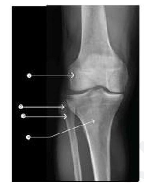
Question 517: Injury at which of the following marked sites on the leg causes failure of dorsiflexion?
- A. Anterior aspect of the thigh (site 1)
- B. Medial aspect of the leg (site 4)
- C. Lateral aspect of the leg (site 3) (Correct Answer)
- D. Posterior aspect of the thigh (site 2)
Explanation: ***Lateral aspect of the leg (site 3)*** - Site 3 points to the **fibula head** and the adjacent region on the lateral aspect of the leg. This is the anatomical location where the **common fibular nerve (peroneal nerve)** wraps around. - The common fibular nerve innervates the muscles responsible for **dorsiflexion** and eversion of the foot. Damage to this nerve, often due to trauma at the fibular neck, leads to **foot drop** and an inability to dorsiflex the foot. *Anterior aspect of the thigh (site 1)* - Site 1 points to the distal femur, which is part of the thigh. Nerves in the anterior thigh (e.g., **femoral nerve**) primarily control hip flexion and knee extension. - Damage here would affect movements of the hip and knee, not directly causing failure of dorsiflexion of the foot. *Medial aspect of the leg (site 4)* - Site 4 points to the medial tibia. This area is associated with the **tibial nerve** and saphenous nerve, which primarily innervate muscles for plantarflexion and inversion of the foot, or provide sensory innervation. - Injury to the tibial nerve would result in an inability to plantarflex and invert the foot, not dorsiflexion. *Posterior aspect of the thigh (site 2)* - Site 2 points to the posterior aspect of the thigh, which is the region for the hamstrings. The **sciatic nerve** and its branches (tibial and common fibular) pass through this region. - While the common fibular nerve originates from the sciatic nerve in the posterior thigh, an injury at this level would likely cause more widespread motor and sensory deficits than isolated dorsiflexion failure, and site 3 is a more common and specific site for common fibular nerve injury isolated to foot drop.
Question 518: Hunter's canal is seen in?
- A. Cubital fossa
- B. Popliteal fossa
- C. Thigh (Correct Answer)
- D. Calf
Explanation: ***Thigh*** - **Hunter's canal**, also known as the **adductor canal**, is an intermuscular passageway located in the **middle third of the thigh**. - It transmits the **femoral artery and vein**, the **saphenous nerve**, and the **nerve to the vastus medialis**. *Cubital fossa* - The **cubital fossa** is a triangular depression located anterior to the elbow joint. - It contains structures like the **brachial artery**, **median nerve**, and biceps tendon, but not Hunter's canal. *Popliteal fossa* - The **popliteal fossa** is a diamond-shaped space located posterior to the knee joint. - It contains the **popliteal artery and vein**, **tibial and common fibular nerves**, and lymph nodes, but not Hunter's canal. *Calf* - The **calf** refers to the posterior compartment of the lower leg. - It houses muscles like the gastrocnemius and soleus, as well as the tibial nerve and posterior tibial artery, but not Hunter's canal.
Question 519: Which artery is primarily responsible for supplying the head and neck of the femur?
- A. Medial circumflex artery (Correct Answer)
- B. Obturator artery
- C. Lateral circumflex artery
- D. Profunda femoris artery
Explanation: ***Medial circumflex artery*** - The **medial circumflex artery** is the primary blood supply to the **femoral head and neck** in adults. - Its branches, particularly the **retinacular arteries**, ascend along the femoral neck to perfuse the head. *Lateral circumflex artery* - The **lateral circumflex artery** supplies the **vastus lateralis muscle** and contributes to the supply of the **greater trochanter**. - While it anastomoses with the medial circumflex artery, its direct contribution to the femoral head is minimal. *Profunda femoris artery* - The **profunda femoris artery**, or deep femoral artery, is the main arterial supply to the **thigh muscles**. - It gives rise to the medial and lateral circumflex femoral arteries but does not directly supply the femoral head. *Obturator artery* - The **obturator artery** primarily supplies the **adductor muscles** of the thigh and contributes branches to the hip joint capsule. - While it has a small branch (artery to the head of the femur) that may contribute to the femoral head in children, it is not the main source in adults.
Question 520: Which is the largest nerve that exits the pelvis through the greater sciatic foramen?
- A. Sciatic nerve (Correct Answer)
- B. Superior gluteal artery
- C. Inferior gluteal artery
- D. Piriformis muscle
Explanation: ***Sciatic nerve*** - The **sciatic nerve** is the largest nerve in the human body, formed from the sacral plexus, and it is indeed the largest structure that passes through the **greater sciatic foramen** as it descends into the posterior thigh. - It supplies motor and sensory innervation to the posterior thigh, lower leg, and foot. *Superior gluteal artery* - The superior gluteal artery exits the pelvis through the **greater sciatic foramen** above the piriformis muscle. - While significant, it is an artery and not a nerve, and it is not the largest structure passing through this foramen. *Inferior gluteal artery* - The inferior gluteal artery also exits the pelvis via the **greater sciatic foramen**, inferior to the piriformis muscle. - Like the superior gluteal artery, it is an arterial structure and not a nerve, and it is not the largest structure in the foramen. *Piriformis muscle* - The **piriformis muscle** originates inside the pelvis and passes through the **greater sciatic foramen** to insert on the greater trochanter of the femur. - Although it occupies a significant portion of the foramen, it is a muscle, not a nerve, and the sciatic nerve is the largest nerve exiting this aperture.
Practice by Chapter
Gluteal Region and Hip
Practice Questions
Thigh and Popliteal Fossa
Practice Questions
Leg and Foot
Practice Questions
Joints of Lower Limb
Practice Questions
Nerves of Lower Limb
Practice Questions
Arterial Supply and Venous Drainage
Practice Questions
Lymphatic Drainage
Practice Questions
Muscles and Their Actions
Practice Questions
Gait Analysis and Biomechanics
Practice Questions
Applied Anatomy and Clinical Correlations
Practice Questions
Want unlimited practice?
Get full access to all questions, explanations, and performance tracking.
Start For Free