
Brain and Neuroanatomy — MCQs

On this page
Infundibular diverticulum is an extension of which structure?
Blood supply of putamen includes all except?
Where is the primary reward center located in the brain?
Which is the only nerve that exits the brainstem dorsally?
What does the cribriform plate form?
A patient is brought to the OPD by his wife, complaining about difficulty expressing emotions and lack of participation in daily activities. On examination, resting tremors and rigidity are noted. Given the possible diagnosis, which part of the brain is affected in this patient?
Which of the following arteries does not supply the medulla?
Identify the marked structure in the image.
Identify the type of the fibre marked in the image of the internal capsule.
A 65-year-old lady presents with a vascular injury to the inferior frontal gyrus. Which functional area would be most affected?
Brain and Neuroanatomy Indian Medical PG Practice Questions and MCQs
Question 431: Infundibular diverticulum is an extension of which structure?
- A. 3rd ventricle (Correct Answer)
- B. 4th ventricle
- C. 1st and 2nd ventricles
- D. None of the options
Explanation: ***3rd ventricle*** * The **infundibular diverticulum** is a small, funnel-shaped extension of the floor of the **third ventricle** of the brain. * It is directly related to the **pituitary gland** and forms the stalk (infundibulum) connecting the hypothalamus to the posterior pituitary. *1st and 2nd ventricles* * The first and second ventricles are the **lateral ventricles**, located within the cerebral hemispheres, and are not directly associated with the infundibular diverticulum. * Their primary connections are to the third ventricle via the **foramina of Monro**. *4th ventricle* * The fourth ventricle is located in the **brainstem**, between the cerebellum and the pons/medulla. * It is connected to the third ventricle by the **aqueduct of Sylvius** and is not the source of the infundibular diverticulum. *None of the options* * This option is incorrect because the infundibular diverticulum is definitively an extension of the **third ventricle**.
Question 432: Blood supply of putamen includes all except?
- A. Lateral striate arteries
- B. Anterior choroidal artery
- C. Medial striate arteries
- D. Posterior communicating artery (Correct Answer)
Explanation: ***Posterior communicating artery*** - The **posterior communicating artery** primarily supplies parts of the **thalamus**, **hypothalamus**, and **midbrain**, but not the putamen. - Its main role is to form part of the **Circle of Willis**, connecting the anterior and posterior cerebral circulations. *Medial striate arteries* - The **medial striate arteries** (or recurrent artery of Heubner) contribute to the blood supply of the **anteromedial part of the globus pallidus** and the **anterior limb of the internal capsule**, as well as parts of the putamen. - They are branches of the **anterior cerebral artery**. *Lateral striate arteries* - The **lateral striate arteries** [1] are crucial for supplying the **putamen**, **globus pallidus**, and the **posterior limb of the internal capsule**. - These arteries are predominantly branches of the **middle cerebral artery**. *Anterior choroidal artery* - The **anterior choroidal artery** supplies the **globus pallidus**, substantial portions of the **internal capsule**, and also contributes to the blood supply of the **posterolateral part of the putamen**. - It arises from the **internal carotid artery**.
Question 433: Where is the primary reward center located in the brain?
- A. Amygdala
- B. Hippocampus
- C. Cerebellum
- D. Nucleus Accumbens (Correct Answer)
Explanation: ***Nucleus Accumbens*** - The **nucleus accumbens** is a crucial part of the **ventral striatum** and is a primary component of the brain's **reward system**. - It plays a central role in processing motivation, pleasure, and reinforcement learning, especially regarding **reward-seeking behaviors**. *Amygdala* - The **amygdala** is primarily involved in processing **emotions**, especially fear and anxiety [2]. - While it interacts with reward pathways, its main role is not as the primary reward center but rather in emotional learning and memory [2]. *Hippocampus* - The **hippocampus** is critical for **memory formation** and spatial navigation [1]. - It plays a role in contextual memory related to rewards but is not the primary site for reward processing itself [1]. *Cerebellum* - The **cerebellum** is largely involved in **motor control**, coordination, balance, and fine-tuning movements. - While it has been implicated in certain cognitive functions, it is not considered part of the brain's primary reward system.
Question 434: Which is the only nerve that exits the brainstem dorsally?
- A. Facial
- B. Trigeminal
- C. Trochlear (Correct Answer)
- D. Abducent
Explanation: ***Trochlear*** - The **trochlear nerve (CN IV)** is unique among cranial nerves as it is the only one that **exits the brainstem dorsally**, specifically from the **dorsal midbrain**. - After exiting dorsally, it then **decussates** (crosses over to the opposite side) before innervating the **superior oblique muscle** of the eye. - It is the **smallest cranial nerve** by number of axons and has the **longest intracranial course**. *Facial* - The **facial nerve (CN VII)** exits the brainstem ventrally, specifically at the **pontomedullary junction**. - It is primarily responsible for **facial expression**, taste from the anterior two-thirds of the tongue, and parasympathetic innervation to glands. *Trigeminal* - The **trigeminal nerve (CN V)** exits the brainstem ventrally from the **lateral aspect of the pons**. - It is the main sensory nerve of the face and also innervates the **muscles of mastication**. *Abducent* - The **abducent nerve (CN VI)** exits the brainstem ventrally, also at the **pontomedullary junction**. - It exclusively innervates the **lateral rectus muscle**, responsible for abducting the eye.
Question 435: What does the cribriform plate form?
- A. Roof of olfactory region (Correct Answer)
- B. Floor of olfactory region
- C. Nasal septum
- D. Lateral wall of nasal cavity
Explanation: The human olfactory epithelium contains about 50 million bipolar olfactory sensory neurons interspersed with glial-like supporting (sustentacular) cells and basal stem cells [1]. ***Roof of olfactory region*** - The **cribriform plate** is a perforated bony plate that forms the **roof of the nasal cavity**, specifically the olfactory region. - Its perforations (foramina) allow the **olfactory nerves (CN I)** to pass from the nasal cavity into the cranial cavity to reach the olfactory bulb. - It is part of the **ethmoid bone** and separates the nasal cavity from the anterior cranial fossa [1]. *Floor of olfactory region* - The **floor of the olfactory region** is primarily formed by the **hard palate** (palatine bone and maxilla). - The cribriform plate is superior to this region, not inferior. *Nasal septum* - The **nasal septum** divides the nasal cavity into two halves, formed by the **vomer**, **perpendicular plate of the ethmoid bone**, and septal cartilage. - While the ethmoid bone contributes to the septum, the cribriform plate specifically forms the roof, not the septum. *Lateral wall of nasal cavity* - The **lateral wall** is formed by several bones including the **maxilla, palatine, inferior concha**, and **medial surface of the ethmoid labyrinth**. - The cribriform plate is a horizontal structure forming the roof, not the lateral wall.
Question 436: A patient is brought to the OPD by his wife, complaining about difficulty expressing emotions and lack of participation in daily activities. On examination, resting tremors and rigidity are noted. Given the possible diagnosis, which part of the brain is affected in this patient?
- A. Basal ganglia (Correct Answer)
- B. Hippocampus
- C. Cerebellum
- D. Premotor cortex
Explanation: **Basal ganglia (Correct)** - The symptoms described—**resting tremors**, **rigidity**, difficulty expressing emotions, and lack of participation—are classic features of **Parkinson's disease**, which is characterized by the degeneration of dopaminergic neurons in the **substantia nigra**, a component of the basal ganglia [1]. - The basal ganglia play a crucial role in motor control, learning, and emotion, and their dysfunction leads to the characteristic motor and non-motor symptoms observed [2]. *Hippocampus (Incorrect)* - The hippocampus is primarily involved in **memory formation** and spatial navigation. - Damage to the hippocampus typically results in **amnesia** or difficulties with new learning, not motor symptoms like tremors or rigidity [3]. *Cerebellum (Incorrect)* - The cerebellum is responsible for **coordination**, balance, and fine motor control [2]. - **Cerebellar dysfunction** typically manifests as **ataxia**, dysmetria, and intention tremors, which differ from the resting tremors and rigidity seen in this patient. *Premotor cortex (Incorrect)* - The premotor cortex is involved in the planning and preparation of movements, as well as the control of trunk and proximal limb muscles. - While it contributes to motor control, its primary dysfunction does not typically cause the combination of **resting tremors** and **rigidity** characteristic of Parkinson's disease.
Question 437: Which of the following arteries does not supply the medulla?
- A. Basilar artery
- B. Anterior spinal artery
- C. Vertebral artery
- D. Posterior cerebral artery (Correct Answer)
Explanation: ***Posterior cerebral artery*** - The **posterior cerebral artery** primarily supplies the **occipital lobe**, the inferior and medial temporal lobes, and parts of the diencephalon and midbrain, not the medulla. - Its territory is typically superior to the medulla's vascular supply. *Basilar artery* - The **basilar artery** is formed by the union of the vertebral arteries and gives rise to several branches that supply the brainstem, including the pons and parts of the medulla. - Branches like the **anterior inferior cerebellar artery (AICA)** and **superior cerebellar artery (SCA)** can have anastomoses that contribute to medullary supply. *Anterior spinal artery* - The **anterior spinal artery**, formed from branches of the vertebral arteries, supplies the anterior two-thirds of the spinal cord and extends rostrally to supply a significant portion of the **medial medulla**. - It is crucial for supplying motor pathways and vital centers in the medulla. *Vertebral artery* - The **vertebral arteries** directly supply the medulla through their branches, including the **posterior inferior cerebellar artery (PICA)** and direct medullary branches. - They also give rise to the anterior and posterior spinal arteries which contribute to medullary supply.
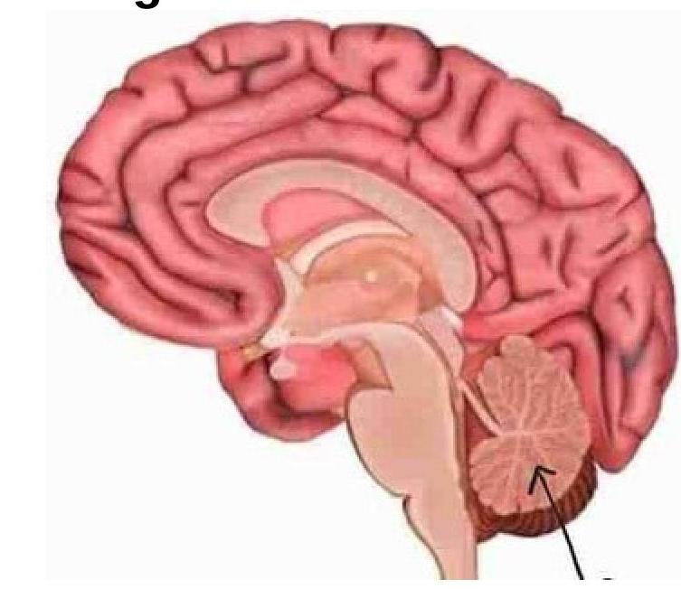
Question 438: Identify the marked structure in the image.
- A. Cerebrum
- B. Brain stem
- C. Corpus callosum
- D. Cerebellum (Correct Answer)
Explanation: ***Cerebellum*** - The image points to the distinct, posterior inferior structure of the brain, characterized by its **folia** and arbour-vitae-like internal structure, which is the cerebellum. - The cerebellum is primarily involved in **motor control**, including coordination, precision, and accurate timing. *Cerebrum* - The cerebrum is the **largest part of the brain**, located superiorly, responsible for higher functions like thought, voluntary movement, and sensory processing. - It consists of two hemispheres connected by the corpus callosum and is characterized by its **gyri** and **sulci**. *Brain stem* - The brain stem is located inferior to the cerebrum and anterior to the cerebellum, connecting the cerebrum and cerebellum to the **spinal cord**. - It controls vital functions such as **breathing**, heart rate, and sleep, and is composed of the midbrain, pons, and medulla oblongata. *Corpus callosum* - The corpus callosum is a large, C-shaped nerve fiber bundle located deep within the brain, under the cerebral cortex. - Its primary function is to **connect the two cerebral hemispheres**, facilitating communication between them.
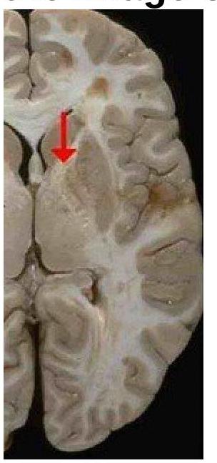
Question 439: Identify the type of the fibre marked in the image of the internal capsule.
- A. Projection fibers (Correct Answer)
- B. Short association fibers
- C. Long association fibers
- D. Commissural fibers
Explanation: ***Projection fibers*** - The image shows the **internal capsule**, which is a white matter structure composed of **projection fibers** that connect the cerebral cortex to subcortical structures, brainstem, and spinal cord. - These fibers facilitate communication between different levels of the central nervous system, including motor and sensory pathways. *Short association fibers* - These fibers, also known as **U-fibers**, connect adjacent gyri within the **same cerebral hemisphere**. - They are typically located superficially in the cerebral cortex, not deep within the brain as shown in the internal capsule. *Long association fibers* - These fibers connect **different lobes** within the **same cerebral hemisphere**, such as the arcuate fasciculus connecting temporal and frontal lobes. - While they are white matter tracts, they do not constitute the internal capsule, which is specifically known for its extensive projection pathways. *Commissural fibers* - **Commissural fibers** connect corresponding areas in the **two cerebral hemispheres**, with the most prominent example being the **corpus callosum**. - The internal capsule, shown in the image, primarily consists of fibers projecting superiorly and inferiorly, rather than horizontally across hemispheres.
Question 440: A 65-year-old lady presents with a vascular injury to the inferior frontal gyrus. Which functional area would be most affected?
- A. Visual
- B. Wernicke
- C. Motor speech (Correct Answer)
- D. Auditory
Explanation: ***Motor speech*** - The **inferior frontal gyrus** is home to **Broca's area**, which is critically involved in **motor speech production** [1]. - A vascular injury here would lead to **expressive aphasia**, where the ability to produce coherent speech is impaired despite intact comprehension [1]. *Visual* - The **visual cortex** is primarily located in the **occipital lobe**, which is at the posterior part of the brain, not the frontal gyrus. - Damage to this area would affect vision, potentially causing **hemianopia** or **cortical blindness**. *Auditory* - The **auditory cortex** is found in the **temporal lobe**, specifically the **superior temporal gyrus** [1]. - Injury to this region would impair the processing of sounds and potentially lead to forms of **auditory agnosia** [2]. *Wernicke* - **Wernicke's area**, responsible for **language comprehension**, is typically located in the **posterior part of the superior temporal gyrus**, in the temporal lobe [1]. - Damage to Wernicke's area results in **receptive aphasia**, where speech comprehension is affected, but speech production remains fluent though often nonsensical [1].
Practice by Chapter
Cerebral Hemispheres
Practice Questions
Diencephalon
Practice Questions
Brainstem
Practice Questions
Cerebellum
Practice Questions
Basal Ganglia
Practice Questions
Limbic System
Practice Questions
Ventricular System and CSF
Practice Questions
Blood Supply of the Brain
Practice Questions
Cranial Nerves and Nuclei
Practice Questions
Functional Systems and Pathways
Practice Questions
Applied Neuroanatomy
Practice Questions
Neuroimaging Correlations
Practice Questions
Want unlimited practice?
Get full access to all questions, explanations, and performance tracking.
Start For Free