Liver Tumors Indian Medical PG Practice Questions and MCQs
Practice Indian Medical PG questions for Liver Tumors. These multiple choice questions (MCQs) cover important concepts and help you prepare for your exams.
Liver Tumors Indian Medical PG Question 1: What is the next best step for a 22-year-old with a hepatic hemangioma on ultrasound?
- A. Angiography
- B. CT
- C. Biopsy
- D. MRI (Correct Answer)
Liver Tumors Explanation: ***MRI***
- **Magnetic Resonance Imaging (MRI)** is the most sensitive and specific imaging modality for confirming the diagnosis of a **hepatic hemangioma** due to its characteristic enhancement patterns.
- An MRI with contrast (e.g., gadolinium) can definitively distinguish a hemangioma from other **benign or malignant liver lesions**, especially when the ultrasound findings are equivocal.
*Angiography*
- **Angiography** is an invasive procedure and is typically reserved for cases where **embolization** or surgical resection of a very large or symptomatic hemangioma is being considered.
- It is not the initial diagnostic choice for confirming a suspected hemangioma identified on **ultrasound**.
*CT*
- A **CT scan** with contrast can also characterize a hemangioma, showing peripheral nodular enhancement followed by progressive centripetal fill-in.
- However, **MRI** generally offers superior soft tissue contrast and provides more definitive diagnostic features for hemangiomas, particularly in younger patients where radiation exposure from CT is a concern.
*Biopsy*
- **Biopsy** of a suspected hepatic hemangioma is generally contraindicated due to the risk of **hemorrhage** and is rarely necessary for diagnosis.
- Imaging characteristics (especially on MRI) are usually sufficient to confirm the diagnosis without the need for an invasive procedure.
Liver Tumors Indian Medical PG Question 2: Skeletal metastasis is common in:
- A. Hepatoma
- B. Cancer stomach
- C. Cancer pancreas
- D. Cancer breast (Correct Answer)
Liver Tumors Explanation: ***Cancer breast***
- **Breast cancer** is one of the most common primary malignancies that metastasize to bones (part of the classic **"osteophilic pentad"**: breast, prostate, lung, kidney, and thyroid) [1].
- Preferentially metastasizes to **axial skeleton** including the **spine**, **pelvis**, and **ribs**.
- Bone metastases from breast cancer can be **osteolytic**, **osteoblastic**, or mixed, often causing pain and **pathological fractures**.
- Approximately **70% of patients with advanced breast cancer** develop bone metastases.
*Hepatoma*
- **Hepatocellular carcinoma (HCC)**, or hepatoma, commonly metastasizes to the **lungs** and regional lymph nodes via hematogenous spread [2].
- While bone metastases can occur, they are **uncommon** (occurs in <5% of cases), much less frequent than with breast, prostate, or lung cancers [1].
- Bone involvement often indicates advanced disease.
*Cancer stomach*
- **Gastric cancer** primarily metastasizes to nearby **lymph nodes**, the **liver**, and the **peritoneum** (Krukenberg tumor to ovaries, Sister Mary Joseph nodule).
- Bone metastases from gastric cancer are **relatively rare**, occurring in less than 10-15% of cases, and represent a poor prognostic sign.
- Not among the common cancers with bone tropism [1].
*Cancer pancreas*
- **Pancreatic cancer** frequently metastasizes to the **liver** (most common), **peritoneum**, and **lungs**.
- Bone metastases are **uncommon** in pancreatic cancer (<5% of cases), typically indicating widespread disease and a very poor prognosis.
- Not part of the osteophilic group of cancers [1].
**References:**
[1] Cross SS. Underwood's Pathology: A Clinical Approach. 6th ed. Common Clinical Problems From Osteoarticular And Connective Tissue Disease, pp. 671-672.
[2] Kumar V, Abbas AK, et al.. Robbins and Cotran Pathologic Basis of Disease. 9th ed. Neoplasia, p. 282.
Liver Tumors Indian Medical PG Question 3: Most common benign tumor of the liver is:
- A. Focal nodular hyperplasia (FNH)
- B. Hepatic adenoma
- C. Hepatic hemangioma (Correct Answer)
- D. Angiomyolipoma of the liver
Liver Tumors Explanation: ***Hepatic hemangioma***
- **Hepatic hemangiomas** are the **most common benign solid tumors of the liver**, often discovered incidentally [1].
- They are composed of a tangled mass of **blood vessels** and are generally asymptomatic [1].
*Focal nodular hyperplasia (FNH)*
- FNH is the **second most common benign liver tumor**, characterized by a central scar on imaging [1].
- While benign, it is less common than hepatic hemangioma [1].
*Hepatic adenoma*
- Hepatic adenomas are benign tumors with a higher risk of **hemorrhage** and **malignant transformation** compared to hemangiomas [1], [2].
- Their incidence is linked to oral contraceptive use or anabolic steroid use.
*Angiolipoma of the liver*
- **Angiomyolipomas** are rare benign tumors, more commonly found in the kidney, and are not the most frequent benign liver tumor.
- They are composed of varying amounts of **fat**, **smooth muscle**, and **blood vessels**.
**References:**
[1] Cross SS. Underwood's Pathology: A Clinical Approach. 6th ed. Common Clinical Problems From Liver And Biliary System Disease, pp. 398-399.
[2] Kumar V, Abbas AK, et al.. Robbins and Cotran Pathologic Basis of Disease. 9th ed. Liver and Gallbladder, pp. 874-875.
Liver Tumors Indian Medical PG Question 4: A patient with a history of alcohol dependence syndrome presents with sudden and unintentional weight loss. What is the most likely diagnosis?
- A. Hepatic adenoma
- B. Cholangiocarcinoma
- C. Hepatocellular carcinoma (Correct Answer)
- D. Alcoholic hepatitis
Liver Tumors Explanation: ***Hepatocellular carcinoma***
- The **alpha-fetoprotein (AFP)** level of **600 ng/mL** is significantly elevated, suggesting a diagnosis of hepatocellular carcinoma, especially in a patient with a history of **alcohol dependence syndrome** [1].
- The **AST/ALT ratio of 0.5** indicates significant liver damage, commonly seen in chronic liver disease leading to **hepatocellular cancer**.
*Alcoholic hepatitis*
- Typically presents with **elevated AST and ALT**, usually with a ratio >2:1, which is not the case here [2].
- May cause weight loss, as alcoholic patients often lose weight due to self-neglect and poor dietary intake, but the **elevated AFP** is not characteristic of merely alcoholic hepatitis [3].
*Cholangiocarcinoma*
- This type of cancer primarily presents with **biliary obstruction** symptoms, such as jaundice, which is not indicated here given **normal bilirubin levels**.
- Does not typically lead to such high levels of **AFP**, making it less likely with the provided lab results.
*Hepatic adenoma*
- More commonly associated with **oral contraceptive use** or anabolic steroid use, not primarily alcohol dependence.
- AFP levels are usually normal or only mildly elevated in hepatic adenoma, making this option less viable with an **AFP level of 600 ng/mL**.
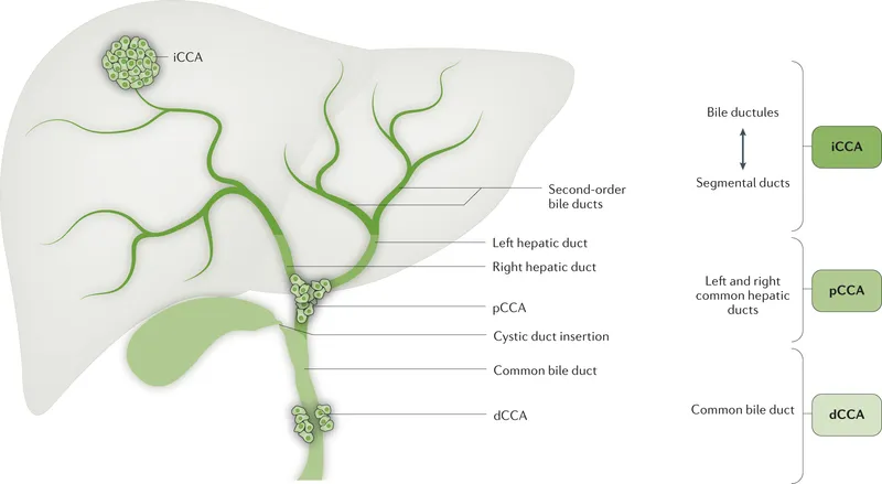
Liver Tumors Indian Medical PG Question 5: What is a Klatskin tumor?
- A. Fibrolamellar hepatocellular carcinoma
- B. Gall bladder carcinoma
- C. Hepatocellular carcinoma
- D. Hilar cholangiocarcinoma (Correct Answer)
Liver Tumors Explanation: ***Nodular type of cholangiocarcinoma***
- Klatskin tumors are a specific form of **cholangiocarcinoma** occurring at the junction of the left and right hepatic bile ducts [1].
- These tumors are characterized by **biliary obstruction** and often present with **jaundice** as a prominent clinical feature.
*Fibrolamellar hepatocellular carcinoma*
- This is a variant of **hepatocellular carcinoma** known for its fibrous stroma, distinct from Klatskin tumors which arise from bile ducts.
- **Fibrolamellar** is more common in younger patients and typically does not cause **biliary obstruction** characteristic of Klatskin tumors.
*Gall bladder carcinoma*
- Gall bladder carcinoma originates from the **gallbladder epithelium**, not the bile ducts, differentiating it from Klatskin tumors.
- It may present with symptoms such as **abdominal pain** and **weight loss**, rather than the specific obstructive jaundice seen in Klatskin cases.
*Hepatocellular carcinoma*
- This cancer arises directly from hepatocytes and is unrelated to bile duct tumors like Klatskin tumors.
- Commonly linked to **chronic liver disease** and liver cirrhosis, it does not typically present with **obstructive jaundice** as seen in cholangiocarcinomas [1].
**References:**
[1] Kumar V, Abbas AK, et al.. Robbins and Cotran Pathologic Basis of Disease. 9th ed. Liver and Gallbladder, pp. 880-881.
Liver Tumors Indian Medical PG Question 6: A patient presents with multiple secondary types of lesions in the liver, non-responding diarrhea, and flushing. It is most probably due to a lesion in:
- A. Liver
- B. Stomach
- C. Small intestine (Correct Answer)
- D. Large intestine
Liver Tumors Explanation: ***Small intestine***
- The presence of **non-responding diarrhea** and **flushing** suggests a neuroendocrine tumor, particularly one that secretes **somatostatin**, commonly associated with the small intestine [1].
- The **multiple liver lesions** often indicate metastasis from an intestinal primary, further supporting a lesion in the small intestine [1].
*Liver*
- While liver lesions are present, they are **secondary** and not the primary source of symptoms, which are more indicative of a gastrointestinal origin [1].
- Liver lesions alone do not typically cause **flushing** or **non-responding diarrhea** without an associated primary tumor in the intestine.
*Stomach*
- Gastric lesions can cause various symptoms but are less commonly associated with **flushing** and **diarrhea**, particularly of the non-responsive type seen here.
- Stomach-specific conditions often relate more to **ulcerations** or bleeding than the systemic endocrine symptoms presented.
*Large intestine*
- While lesions in the large intestine can lead to **diarrhea**, they do not often present with **flushing** or involve multiple liver lesions.
- This type of presentation is more characteristic of small intestinal involvement, especially in cases of **carcinoid syndrome** from neuroendocrine tumors [1].
Liver Tumors Indian Medical PG Question 7: Which of the following statements about hepatocellular carcinoma (HCC) is true?
- A. It is resectable in early stages, depending on size, location, and liver function.
- B. More than 70% of cases show increased AFP levels.
- C. It is the most common primary liver tumor, though metastatic tumors are more common overall.
- D. All of the options. (Correct Answer)
Liver Tumors Explanation: ***All of the options.***
- This option is correct because HCC is indeed resectable in early stages under specific conditions, AFP levels are elevated in a significant portion of cases, and it is the most common primary liver tumor. [1]
- Each individual statement provides an accurate insight into the characteristics and clinical aspects of hepatocellular carcinoma. [2]
*It is resectable in early stages, depending on size, location, and liver function.*
- **Early-stage HCC** can be potentially cured with **surgical resection** or liver transplantation, provided the tumor is small, solitary, and the patient has good liver function. [2]
- The decision for resectability is complex and depends on factors such as **tumor size, location**, and the patient's **Child-Pugh score** or MELD score for liver function.
*More than 70% of cases show increased AFP levels.*
- **Alpha-fetoprotein (AFP)** is a widely used **tumor marker** for HCC, and elevated levels are observed in 60-80% of patients, particularly those with larger tumors. [1]
- While helpful for screening and monitoring, AFP is not specific enough for diagnosis on its own, as it can also be elevated in other liver conditions and germ cell tumors.
*It is the most common primary liver tumor, though metastatic tumors are more common overall.*
- **Hepatocellular carcinoma (HCC)** accounts for about 80-90% of **primary liver cancers**, making it the most prevalent type originating in the liver.
- However, **metastatic liver cancer**, meaning cancer that has spread to the liver from another primary site (e.g., colon, lung, breast), is significantly more common in the general population than primary liver cancers.
Liver Tumors Indian Medical PG Question 8: Statement 1 - A 59-year-old patient presents with flaccid bullae. Histopathology shows a suprabasal acantholytic split.
Statement 2 - The row of tombstones appearance is diagnostic of Pemphigus vulgaris.
- A. Statements 1 & 2 are correct, 2 is not explaining 1 (Correct Answer)
- B. Statements 1 and 2 are correct and 2 is the correct explanation for 1
- C. Statements 1 and 2 are incorrect
- D. Statement 1 is incorrect
Liver Tumors Explanation: ***Correct: Statements 1 & 2 are correct, 2 is not explaining 1***
**Analysis of Statement 1:**
- A 59-year-old patient with **flaccid bullae** and **suprabasal acantholytic split** on histopathology is the classic presentation of **Pemphigus vulgaris**
- The flaccid (easily ruptured) nature of bullae distinguishes it from tense bullae seen in bullous pemphigoid
- The suprabasal location of the split (just above the basal layer) with acantholysis (loss of cell-to-cell adhesion) is pathognomonic
- **Statement 1 is CORRECT** ✓
**Analysis of Statement 2:**
- The **"row of tombstones" or "tombstone appearance"** is indeed a diagnostic histopathological feature of Pemphigus vulgaris
- This appearance results from basal keratinocytes remaining attached to the basement membrane while suprabasal cells separate due to acantholysis
- The intact basal cells standing upright resemble a row of tombstones
- **Statement 2 is CORRECT** ✓
**Does Statement 2 explain Statement 1?**
- Statement 2 describes a **histopathological appearance** (tombstone pattern) that is a **consequence** of the suprabasal split
- However, it does NOT explain the **underlying cause** of the flaccid bullae or the suprabasal split
- The true explanation involves **IgG autoantibodies against desmoglein 3 (and desmoglein 1)**, which attack intercellular adhesion structures (desmosomes), causing **acantholysis**
- Therefore, **Statement 2 does NOT explain Statement 1** ✗
*Incorrect: Statement 2 is the correct explanation for Statement 1*
- While both statements describe features of Pemphigus vulgaris, the tombstone appearance is a descriptive finding, not an explanatory mechanism
*Incorrect: Statements 1 and 2 are incorrect*
- Both statements are medically accurate descriptions of Pemphigus vulgaris features
*Incorrect: Statement 1 is incorrect*
- Statement 1 correctly describes the cardinal clinical and histopathological features of Pemphigus vulgaris
Liver Tumors Indian Medical PG Question 9: Which of the following liver tumors has a propensity to invade the portal or hepatic vein?
- A. Cavernous hemangioma
- B. Hepatocellular carcinoma (Correct Answer)
- C. Focal nodular hyperplasia
- D. Hepatic adenoma
Liver Tumors Explanation: ***Hepatocellular carcinoma*** - **Hepatocellular carcinoma (HCC)** is known for its aggressive nature and a characteristic tendency to invade vascular structures, particularly the **portal vein** or hepatic veins [1]. - This **vascular invasion** contributes to its metastatic potential and is a critical factor in prognosis and treatment planning [1].*Cavernous hemangioma* - A **cavernous hemangioma** is a benign vascular tumor of the liver, typically recognized as an incidental finding. - While it is a vascular lesion, it does not invade the large hepatic or portal veins but rather consists of **dilated vascular spaces** within the liver parenchyma.*Focal nodular hyperplasia* - **Focal nodular hyperplasia (FNH)** is a **benign liver lesion** characterized by a central fibrous scar and radiating septa [2]. - It is typically well-circumscribed and does not exhibit aggressive features like **vascular invasion** [2].*Hepatic adenoma* - A **hepatic adenoma** is a benign tumor, often associated with oral contraceptive use, which can sometimes pose a risk of rupture or malignant transformation. - However, it does not typically show features of **vascular invasion** into the portal or hepatic veins [2].
**References:**
[1] Kumar V, Abbas AK, et al.. Robbins and Cotran Pathologic Basis of Disease. 9th ed. Liver and Gallbladder, pp. 878-879.
[2] Cross SS. Underwood's Pathology: A Clinical Approach. 6th ed. Common Clinical Problems From Liver And Biliary System Disease, pp. 398-399.
Liver Tumors Indian Medical PG Question 10: Which is a hormone dependent liver tumor?
- A. Adenoma (Correct Answer)
- B. Hemangioma
- C. Hepatocellular carcinoma
- D. Hemangiopericytoma
Liver Tumors Explanation: ***Adenoma***
- Hepatic adenomas are **hormone-dependent tumors** commonly associated with conditions like **oral contraceptive use** and are influenced by estrogen [1].
- These tumors can present as **benign liver masses**, but they have a risk of hemorrhage and malignant transformation [1].
*Hepatocellular carcinoma*
- This is a **malignant tumor** of the liver primarily associated with cirrhosis and chronic liver disease, not directly hormone-dependent.
- Risk factors include **viral hepatitis** and **alcohol exposure**, rather than hormonal influences.
*Hemangioma*
- Liver hemangiomas are **vascular lesions** that are usually asymptomatic and are **not hormone-dependent**.
- They are the most common benign liver tumors, often discovered incidentally during imaging.
*Hemangiopericytoma*
- A rare tumor, hemangiopericytoma originates from **pericytes** around blood vessels and is not specifically associated with liver tissue or hormones.
- It can arise in various organs but lacks the dependency on hormones seen in hepatic adenomas.
**References:**
[1] Kumar V, Abbas AK, et al.. Robbins and Cotran Pathologic Basis of Disease. 9th ed. Liver and Gallbladder, p. 874.
More Liver Tumors Indian Medical PG questions available in the OnCourse app. Practice MCQs, flashcards, and get detailed explanations.