Osteomyelitis Indian Medical PG Practice Questions and MCQs
Practice Indian Medical PG questions for Osteomyelitis. These multiple choice questions (MCQs) cover important concepts and help you prepare for your exams.
Osteomyelitis Indian Medical PG Question 1: A boy presented with multiple non suppurative osteomyelitis with sickle cell anaemia. What will be the causative organism?
- A. Salmonella (Correct Answer)
- B. H. influenzae
- C. Enterobacter species
- D. Staphylococcus aureus
Osteomyelitis Explanation: ***Salmonella***
- **Salmonella species** are a well-known cause of **osteomyelitis** in patients with **sickle cell anemia**, due to factors like gut mucosal damage and functional asplenia. [1]
- The unique pathophysiology of sickle cell disease, including areas of bone infarction and compromised reticulendothelial system function, predisposes these patients to **Salmonella infections**. [1]
*Staphylococcus aureus*
- While **Staphylococcus aureus** is the most common cause of osteomyelitis in the general population, it is less likely to be the causative organism in patients with **sickle cell anemia** compared to Salmonella.
- Its presence usually indicates other predisposing factors like trauma or prosthetic devices.
*H. influenzae*
- **Haemophilus influenzae** was a common cause of osteomyelitis in children before widespread vaccination but is now rare, especially with routine immunizations.
- It is not specifically associated with a higher risk in patients with **sickle cell disease** for osteomyelitis compared to other pathogens.
*Enterobacter species*
- **Enterobacter species** can cause osteomyelitis, particularly in immunocompromised individuals or following surgery, but they are not uniquely associated with **sickle cell anemia** as a primary cause compared to Salmonella.
- Their involvement in non-suppurative osteomyelitis in this specific patient population is less common.
Osteomyelitis Indian Medical PG Question 2: Which microorganism is the most common cause of pyogenic osteomyelitis?
- A. S. aureus (Correct Answer)
- B. Streptococcus spp.
- C. Corynebacterium spp.
- D. Neisseria gonorrhoeae (gonococcus)
Osteomyelitis Explanation: ***Staph aureus***
- **_Staphylococcus aureus_** is the most frequent cause of **pyogenic osteomyelitis** across all age groups and routes of infection.
- Its ability to adhere to bone, form biofilms, and produce toxins contributes to its prevalence in bone infections.
*Streptococcus spp.*
- While various **_Streptococcus_** species can cause infections, they are less common causes of pyogenic osteomyelitis compared to **_Staphylococcus aureus_**.
- **Group A _Streptococcus_** can cause severe invasive infections but rarely involves primary bone infection.
*Corynebacterium spp.*
- **_Corynebacterium_** species, particularly **_Corynebacterium striatum_**, are increasingly recognized as opportunistic pathogens, especially in immunocompromised individuals or those with foreign bodies.
- However, they are not the most common cause of osteomyelitis in the general population.
*Neisseria gonorrhoeae (gonococcus)*
- **_Neisseria gonorrhoeae_** can cause **disseminated gonococcal infection (DGI)**, which may include joint involvement (**septic arthritis**).
- While it can lead to bone pain and swelling, it primarily affects joints and is a less common cause of direct **pyogenic osteomyelitis** than **_S. aureus_**.
Osteomyelitis Indian Medical PG Question 3: 12 years male came with swelling of lower end tibia which is surrounded by rim of reactive bone. What is most likely diagnosis?
- A. GCT
- B. Hyper PTH
- C. Brodie's Abscess (Correct Answer)
- D. Osteomyelitis
Osteomyelitis Explanation: ***Brodie's Abscess***
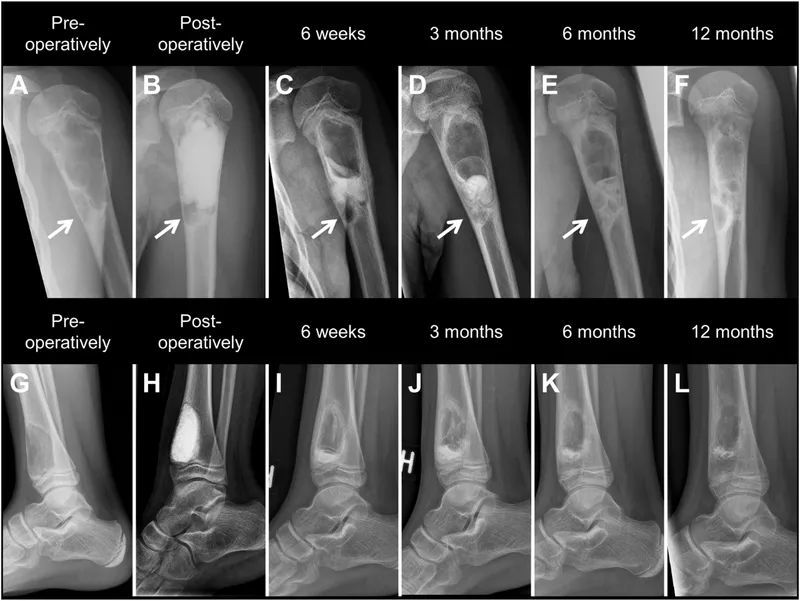
- A **Brodie's abscess** is a subacute or chronic osteomyelitis characterized by a localized bone abscess, typically with a surrounding **sclerotic rim of reactive bone**.
- It often occurs in the **metaphysis of long bones** (like the lower end of the tibia) in children and adolescents, presenting with localized pain and swelling.
*GCT*
- **Giant cell tumor (GCT)** typically occurs in **skeletally mature adults** (20-40 years old) and is a lytic lesion often found in the **epiphysis** of long bones, rarely with a distinct sclerotic rim.
- GCTs are generally more aggressive and demonstrate a **soap-bubble appearance** with cortical expansion rather than a thick reactive bone rim.
*Hyper PTH*
- **Hyperparathyroidism** causes bone changes such as **osteopenia**, **subperiosteal bone resorption**, especially in the phalanges, and **brown tumors** (lytic lesions).
- It does not typically present as a localized lesion with a **sclerotic rim of reactive bone** in a child.
*Osteomyelitis*
- While chronic osteomyelitis can involve local bone destruction and reactive bone formation, a **Brodie's abscess** is a specific, well-circumscribed form of **subacute osteomyelitis**.
- Acute osteomyelitis presents with more diffuse systemic symptoms (fever, malaise) and less defined reactive bone in its early stages compared to the distinct **sclerotic rim** seen in a Brodie's abscess.
Osteomyelitis Indian Medical PG Question 4: A young girl presented with swelling of right thigh, with history of trauma 2 months back. Now she presents with swelling at mid-shaft of femur & low grade fever. ESR is mildly raised. X-ray shows a laminated periosteal reaction. Next line of investigation would be:
- A. MRI (Correct Answer)
- B. Bone scan
- C. Blood count & CRP
- D. Biopsy
Osteomyelitis Explanation: ***MRI***
- An **MRI** is the most appropriate next step as it provides excellent detailed imaging of soft tissues and bone marrow, allowing better characterization of the **periosteal reaction**, identifying abscesses, and assessing the extent of bone involvement, crucial for differentiating between infection and tumor.
- The presence of a **laminated periosteal reaction** (like an "onion peel") on X-ray, in conjunction with localized swelling and low-grade fever, is highly suggestive of subacute osteomyelitis or even some bone tumors like Ewing sarcoma, for which MRI is superior for defining the extent.
*Bone scan*
- A **bone scan** (technetium-99m) is sensitive for detecting increased bone turnover, which occurs in infections and tumors, but it is **non-specific**, meaning it cannot differentiate between these conditions.
- While it could show increased uptake in the affected area, it would not provide the anatomical detail needed to characterize the lesion or guide further management as effectively as an MRI.
*Blood count & CRP*
- A **blood count and CRP** would provide information on systemic inflammation (e.g., leukocytosis, elevated CRP for infection), but these are **non-specific**.
- While ESR is already mildly raised, these blood tests would confirm generalized inflammation but **cannot localize or characterize the lesion** in the bone, offering little diagnostic value for the specific problem at this stage without imaging.
*Biopsy*
- A **biopsy** is an invasive procedure and is typically performed after initial imaging studies like X-ray and MRI have characterized the lesion to guide the biopsy site and help determine the nature of the condition (e.g., infection vs. tumor).
- Performing a biopsy as the immediate next step without detailed imaging to assess the extent and nature of the lesion would be premature and potentially less effective in diagnosis.
Osteomyelitis Indian Medical PG Question 5: The earliest radiological change to appear in a case of acute osteomyelitis is:
- A. Periosteal reaction
- B. Sequestrum formation
- C. Bony sclerosis
- D. Loss of plane between soft tissue and muscle (Correct Answer)
Osteomyelitis Explanation: ***Loss of plane between soft tissue and muscle***
- This finding, often seen as **soft tissue swelling** and effacement of fat planes on radiographs, is the **earliest detectable radiographic sign** in acute osteomyelitis, typically appearing within 24-48 hours.
- It reflects the initial inflammatory changes and **edema** in the soft tissues surrounding the infected bone.
*Periosteal reaction*
- This occurs later than soft tissue changes, usually appearing after **7-10 days** of infection, as the periosteum is lifted and new bone formation begins.
- It is a sign of bone irritation and can be seen as linear or lamellated **new bone growth** parallel to the cortex.
*Sequestrum formation*
- A sequestrum is a piece of **devitalized (necrotic) bone** that separates from the healthy bone, a much later complication of osteomyelitis.
- It typically appears several weeks into the disease course, indicating established bone necrosis and usually requiring surgical intervention.
*Bony sclerosis*
- **Bony sclerosis**, or increased bone density, is a sign of chronic inflammation and new bone formation in response to persistent infection.
- This change is usually observed in the **later stages of osteomyelitis** or in chronic forms, not in the acute phase.
Osteomyelitis Indian Medical PG Question 6: Which of the following statements about osteomyelitis is incorrect?
- A. Sequestrum is a piece of dead bone
- B. Epiphysis most commonly involved region (Correct Answer)
- C. In sickle cell anemia salmonella is causative organism
- D. Involucrum is dense sclerotic bone overlying a sequestrum
Osteomyelitis Explanation: ***Epiphysis most commonly involved region***
- This statement is **incorrect** because osteomyelitis, particularly in children and adolescents, most commonly affects the **metaphysis** of long bones due to its rich, slow-flowing blood supply, which facilitates bacterial deposition.
- The epiphysis is less commonly involved primarily due to the differences in vascularity and growth plate anatomy.
*Sequestrum is a piece of dead bone*
- This statement is **correct**. A **sequestrum** refers to a piece of dead or necrotic bone that has separated from the surrounding healthy bone, often seen in chronic osteomyelitis [1].
- It results from the inflammatory process and lack of blood supply, acting as a nidus for infection.
*Involucrum is dense sclerotic bone overlying a sequestrum*
- This statement is **correct**. An **involucrum** is a new shell of dense, sclerotic bone that forms around a sequestrum in chronic osteomyelitis, attempting to wall off the infection [1].
- It represents the body's attempt to heal and contain the infection, often leading to sinus tract formation [1].
*In sickle cell anemia salmonella is causative organism*
- This statement is **correct**. Patients with **sickle cell anemia** are particularly susceptible to **Salmonella osteomyelitis**, which replaces Staphylococcus aureus as the predominant causative agent in this population.
- The altered splenic function and compromised immune response in sickle cell disease contribute to this increased risk.
**References:**
[1] Kumar V, Abbas AK, et al.. Robbins and Cotran Pathologic Basis of Disease. 9th ed. Bones, Joints, and Soft Tissue Tumors, pp. 1197-1198.
Osteomyelitis Indian Medical PG Question 7: A 25-year-old male presents with localized pain in the tibia and swelling. Imaging reveals a bone abscess. Identify the condition.
- A. Brodie abscess (Correct Answer)
- B. Osteoid osteoma
- C. Intracortical hemangioma
- D. Chondromyxoid fibroma
Osteomyelitis Explanation: ***Brodie abscess***
- A Brodie abscess is a **subacute or chronic osteomyelitis** characterized by a well-circumscribed, **radiolucent lesion** (an abscess cavity) often surrounded by a zone of **sclerosis**, representing the body's attempt to wall off the infection.
- The presentation of localized pain and swelling in the tibia, with imaging revealing a bone abscess, is consistent with this condition, which is a common form of localized osteomyelitis.
*Osteoid osteoma*
- This is a **benign bone tumor** characterized by a small, radiolucent nidus surrounded by a large area of **sclerotic bone**. The pain from an osteoid osteoma is typically **worse at night** and dramatically relieved by NSAIDs.
- While it can cause localized pain and swelling, the imaging features of a distinct abscess cavity are not characteristic of an osteoid osteoma.
*Intracortical hemangioma*
- An intracortical hemangioma is a **rare benign vascular lesion** within the cortex of a bone.
- Imaging typically shows a **lytic lesion** with a characteristic **"honeycomb" or "sunburst" appearance**, not a well-defined abscess.
*Chondromyxoid fibroma*
- This is a rare, **benign cartilaginous tumor** that usually presents as an **eccentric lytic lesion** in the metaphysis of long bones, often with a scalloped border and sclerotic rim.
- While it can cause localized pain and swelling, the imaging appearance of an abscess with sclerotic margins is not typical of a chondromyxoid fibroma.
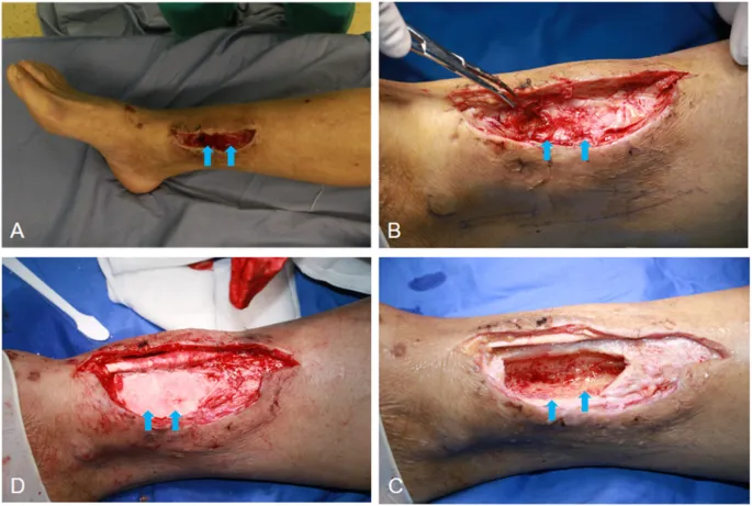
Osteomyelitis Indian Medical PG Question 8: A 28-year-old male with a history of trauma presents with a non-healing sinus on the tibia. An X-ray shows a sequestrum. What is the appropriate next step in management?
- A. Systemic antibiotics
- B. Local wound care
- C. Sequestrectomy (Correct Answer)
- D. Bone grafting
Osteomyelitis Explanation: ***Sequestrectomy***
- A **sequestrum** is a piece of dead bone that has become separated from the surrounding healthy bone during necrosis. In the context of **chronic osteomyelitis**, this dead bone acts as a nidus for infection that cannot be eradicated by antibiotics alone.
- The presence of a **non-healing sinus** and a sequestrum on X-ray clearly indicates **chronic osteomyelitis**, which requires surgical removal of the infected dead bone (sequestrectomy) for resolution.
*Systemic antibiotics*
- While systemic antibiotics are crucial in treating acute osteomyelitis and as an adjunct in chronic cases, they are unlikely to cure an infection with a sequestered dead bone.
- The **avascular nature of the sequestrum** prevents adequate penetration of antibiotics, making them ineffective as a sole therapy.
*Local wound care*
- Local wound care might help manage the non-healing sinus superficially but does not address the underlying **bone infection and dead bone**, which is the primary pathology.
- This approach would only provide symptomatic relief without resolving the infectious process.
*Bone grafting*
- Bone grafting is typically performed after the infection has been completely eradicated and involves filling a bone defect.
- Performing bone grafting while a **sequestrum and ongoing infection** are present would likely lead to graft failure and continued infection.
Osteomyelitis Indian Medical PG Question 9: Which of the following statements about tubercular osteomyelitis is NOT true?
- A. Sequestrum is uncommon
- B. It is a type of secondary osteomyelitis
- C. Periosteal reaction is characteristic (Correct Answer)
- D. Inflammation is minimal
Osteomyelitis Explanation: ***Periosteal reaction is characteristic***
- This statement is **NOT true** for tubercular osteomyelitis; periosteal reaction is generally **minimal or absent** due to the insidious and less florid inflammatory response.
- Unlike pyogenic osteomyelitis, which causes significant periosteal new bone formation, tuberculosis typically results in **slow bone destruction** without marked reactive bone changes.
*Sequestrum is uncommon*
- This statement is **true** because **sequestrum** (a piece of dead bone separated from healthy bone) is less frequently observed in tubercular osteomyelitis compared to pyogenic osteomyelitis.
- The **granulomatous inflammation** of tuberculosis tends to cause slow bone necrosis rather than the rapid, liquefactive necrosis that leads to large sequestra.
*It is a type of secondary osteomyelitis*
- This statement is **true** as tubercular osteomyelitis is almost always secondary to a **primary focus of tuberculosis** elsewhere in the body, typically the lungs [1].
- The infection spreads **hematogenously** to the bone, making it a manifestation of disseminated tuberculosis rather than a primary bone infection [1].
*Inflammation is minimal*
- This statement is **true** in the sense that the **acute inflammatory response** in tubercular osteomyelitis is often less pronounced than in pyogenic infections.
- While it is a chronic infectious process, the characteristic **granulomatous inflammation** develops over time, and the initial or acute inflammatory signs might be subtle or "minimal" compared to bacterial osteomyelitis [1].
Osteomyelitis Indian Medical PG Question 10: A 30 year old previously healthy man presented to the emergency department immediately after being involved in a road traffic accident. After clinical examination, scaphoid injury was suspected. A radiograph of the left wrist was obtained and found to be equivocal. What is the best next step?
- A. MRI Scan
- B. Presumptive Casting (Correct Answer)
- C. Bone scintigraphy of wrist
- D. CT Scan
Osteomyelitis Explanation: ***Presumptive Casting***
- When scaphoid fracture is suspected clinically but **radiographs are equivocal**, conservative management with **presumptive casting** is appropriate.
- This prevents potential avascular necrosis and allows for healing if a fracture is present but not yet visible.
*MRI Scan*
- While an **MRI** is highly sensitive for detecting scaphoid fractures, it is not always immediately available or cost-effective as the very first step following equivocal X-rays in a stable patient.
- Delaying immobilization to obtain an immediate MRI could lead to further displacement or complications if a fracture is indeed present.
*Bone scintigraphy of wrist*
- **Bone scintigraphy** (bone scan) can detect subtle fractures, but it is not typically performed immediately after injury due to its lower specificity and relatively longer time frame to show changes compared to other modalities like MRI.
- It involves radiation and is usually reserved for cases where MRI is contraindicated or unavailable and earlier imaging was inconclusive.
*CT Scan*
- A **CT scan** is excellent for visualizing cortical bone and complex fractures but is less sensitive than MRI for detecting occult scaphoid fractures or soft tissue injuries.
- It also involves significant radiation exposure, making it a secondary option to MRI or conservative management for initial detection.
More Osteomyelitis Indian Medical PG questions available in the OnCourse app. Practice MCQs, flashcards, and get detailed explanations.