Cerebral Cortex Indian Medical PG Practice Questions and MCQs
Practice Indian Medical PG questions for Cerebral Cortex. These multiple choice questions (MCQs) cover important concepts and help you prepare for your exams.
Cerebral Cortex Indian Medical PG Question 1: Ablation of the somatosensory area I of the cerebral cortex leads to
- A. Loss of tactile localization but not of two point discrimination
- B. Loss of tactile localization and two point discrimination (Correct Answer)
- C. Total loss of pain sensation
- D. Total loss of touch sensation
Cerebral Cortex Explanation: ***Loss of tactile localization and two point discrimination***
- The **somatosensory area I (S1)** is crucial for processing higher-order somatic sensations, including the **discriminative aspects of touch**.
- Its ablation leads to deficits in distinguishing distinct points of touch (**two-point discrimination**) and precisely identifying where touch occurred (**tactile localization**).
*Loss of tactile localization but not of two point discrimination*
- This option incorrectly suggests that **two-point discrimination** would be preserved, which is not the case as S1 is essential for both functions.
- While other cortical areas contribute to sensation, S1 is paramount for fine tactile distinctions.
*Total loss of pain sensation*
- **Pain sensation** is processed across multiple brain regions, including the **thalamus**, **insula**, and **anterior cingulate cortex**, not solely S1.
- S1 primarily processes discriminatory aspects of somatosensation, not the affective component of pain.
*Total loss of touch sensation*
- A total loss of touch sensation implies a complete unawareness of touch, which would require more extensive damage than just S1 ablation, potentially affecting **thalamic pathways** or other cortical regions.
- S1 ablation primarily affects the *quality* and *interpretation* of touch, rather than its complete absence.
Cerebral Cortex Indian Medical PG Question 2: What is the primary symptom associated with a lesion in Wernicke's area?
- A. Inability to understand language
- B. Inability to repeat phrases
- C. Fluent speech with poor comprehension (Correct Answer)
- D. Difficulty in forming sentences
Cerebral Cortex Explanation: ***Fluent speech with poor comprehension***
- A lesion in **Wernicke's area** results in **Wernicke's aphasia**, where the individual can produce speech fluently but the content is often meaningless or nonsensical (word salad). [1]
- The primary characteristic is a profound **difficulty in understanding** spoken and written language, despite intact hearing and vision.
*Inability to understand language*
- While an inability to understand language is a key component of Wernicke's aphasia, the description "fluent speech with poor comprehension" more comprehensively captures the clinical presentation by including the intact though often chaotic speech production.
- This option alone does not fully encompass the unique **dissociation between fluency and comprehension** seen in Wernicke's aphasia.
*Inability to repeat phrases*
- The **inability to repeat phrases** is typically associated with **conduction aphasia**, which results from damage to the **arcuate fasciculus**, the connection between Wernicke's and Broca's areas. [1]
- While repetition can be impaired in Wernicke's aphasia due to poor comprehension, it is not the primary defining symptom differentiating it from other aphasias.
*Difficulty in forming sentences*
- **Difficulty in forming sentences** and producing meaningful speech, often characterized by **non-fluent, effortful speech** and agrammatism, is a hallmark of **Broca's aphasia**. [1]
- Broca's area is responsible for **speech production** and grammatical structure, not language comprehension.
Cerebral Cortex Indian Medical PG Question 3: Brain areas involved with obsessive compulsive disorder include all except:
- A. Head of caudate nucleus
- B. Corpus callosum (Correct Answer)
- C. Orbitofrontal cortex
- D. Basal ganglia
Cerebral Cortex Explanation: ***corpus callosum***
- The **corpus callosum** is primarily involved in **interhemispheric communication**, connecting the two cerebral hemispheres, and is not a core area implicated in the pathophysiology of **OCD**.
- While damage to the corpus callosum can lead to neurological deficits, it is not directly associated with the obsessions and compulsions seen in OCD.
*Head of caudate nucleus*
- The **caudate nucleus**, particularly its head, is part of the **basal ganglia** and is highly implicated in **OCD**, with studies showing abnormal activity and volume.
- It plays a crucial role in **goal-directed behavior** and **habit formation**, which are dysfunctional in OCD.
*Orbitofrontal cortex*
- The **orbitofrontal cortex (OFC)** is consistently identified in neuroimaging studies as being hyperactive in individuals with **OCD**.
- It is involved in **decision-making**, **reward processing**, and **emotional regulation**, contributing to the characteristic symptoms of OCD.
*Basal ganglia*
- The **basal ganglia**, a group of subcortical nuclei including the **caudate nucleus**, **putamen**, and **globus pallidus**, are central to the neurocircuitry of **OCD**.
- This region is critical for **motor control**, **habit learning**, and **executive functions**, and its dysfunction is thought to contribute to the repetitive behaviors and cognitive rigidity seen in OCD.
Cerebral Cortex Indian Medical PG Question 4: What are the effects of a lesion in Brodmann area 22?
- A. Expressive aphasia
- B. Receptive aphasia (Correct Answer)
- C. Poor repetition of language
- D. Poor naming
Cerebral Cortex Explanation: ***Receptive aphasia***
- A lesion in **Brodmann area 22**, specifically in **Wernicke's area**, leads to **receptive aphasia** (Wernicke's aphasia).
- This condition is characterized by **impaired comprehension** of spoken and written language, **fluent but paraphasic speech**, and **poor repetition**.
- This is the most comprehensive answer as it describes the entire clinical syndrome.
*Expressive aphasia*
- **Brodmann areas 44 and 45** (Broca's area) in the frontal lobe are associated with expressive aphasia (Broca's aphasia).
- Patients have good comprehension but struggle to produce fluent speech, with effortful, telegraphic output.
*Poor repetition of language*
- While poor repetition is indeed a feature of Wernicke's aphasia, this option describes only one component of the syndrome rather than the complete clinical picture.
- **Conduction aphasia** (from arcuate fasciculus lesions) is characterized by poor repetition with **relatively preserved** comprehension and fluent speech, distinguishing it from Wernicke's aphasia.
- "Receptive aphasia" is the more complete answer.
*Poor naming*
- Difficulty with naming, or **anomia**, is a common feature across various types of aphasia, including both receptive and expressive aphasia.
- It reflects disruption in language networks involving the **temporal and parietal lobes** but is not specific to Brodmann area 22 lesions.
Cerebral Cortex Indian Medical PG Question 5: Match the following drugs in Column A with their contraindications in Column B.
| Column A | Column B |
| :-- | :-- |
| 1. Morphine | 1. QT prolongation |
| 2. Amiodarone | 2. Thromboembolism |
| 3. Vigabatrin | 3. Pregnancy |
| 4. Estrogen preparations | 4. Head injury |
- A. A-1, B-3, C-2, D-4
- B. A-4, B-1, C-3, D-2 (Correct Answer)
- C. A-3, B-2, C-4, D-1
- D. A-2, B-4, C-1, D-3
Cerebral Cortex Explanation: ***A-4, B-1, C-3, D-2***
- **Morphine** is contraindicated in **head injury** as it can increase intracranial pressure and mask neurological symptoms.
- **Amiodarone** is contraindicated in patients with **QT prolongation** due to its risk of inducing more severe arrhythmias like Torsades de Pointes.
- **Vigabatrin** is contraindicated during **pregnancy** due to its potential for teratogenicity and adverse effects on fetal development.
- **Estrogen preparations** are contraindicated in patients with a history of **thromboembolism** due to their increased risk of blood clot formation.
*A-1, B-3, C-2, D-4*
- This option incorrectly matches **Morphine** with QT prolongation and **Estrogen preparations** with head injury, which are not their primary contraindications.
- It also incorrectly links **Vigabatrin** with thromboembolism and **Amiodarone** with pregnancy.
*A-3, B-2, C-4, D-1*
- This choice incorrectly associates **Morphine** with pregnancy and **Vigabatrin** with head injury, which are not the most critical or direct contraindications.
- It also misaligns **Amiodarone** with thromboembolism and **Estrogen preparations** with QT prolongation.
*A-2, B-4, C-1, D-3*
- This option incorrectly matches **Morphine** with thromboembolism and **Amiodarone** with head injury, which are not their most significant contraindications.
- It also incorrectly links **Vigabatrin** with QT prolongation and **Estrogen preparations** with pregnancy.
Cerebral Cortex Indian Medical PG Question 6: Which of the following Brodmann areas is primarily associated with Broca's motor aphasia?
- A. 44 (Correct Answer)
- B. 4
- C. 22
- D. 17
Cerebral Cortex Explanation: ***Brodmann Area 44***
- **Brodmann area 44**, also known as **Broca's area**, is located in the inferior frontal gyrus and is critical for **speech production**.
- Damage to this area typically leads to **Broca's motor aphasia** (also called expressive aphasia), characterized by **non-fluent speech**, difficulty forming words, and impaired grammar while comprehension remains relatively intact.
*Brodmann Area 4*
- **Brodmann area 4** corresponds to the **primary motor cortex**, responsible for executing voluntary movements.
- While essential for motor control, it is not directly involved in the cognitive aspects of language production that define Broca's aphasia.
*Brodmann Area 22*
- **Brodmann area 22** is primarily associated with **Wernicke's area**, which is located in the superior temporal gyrus and is crucial for **language comprehension**.
- Damage to this area results in **Wernicke's aphasia** (receptive aphasia), characterized by fluent but meaningless speech and impaired comprehension.
*Brodmann Area 17*
- **Brodmann area 17** is the **primary visual cortex**, responsible for processing visual information.
- It plays no direct role in language processing or speech production; damage here would primarily cause visual field deficits.
Cerebral Cortex Indian Medical PG Question 7: Which of the following is not a tributary of the cavernous sinus?
- A. Central vein of retina
- B. Sphenoparietal sinus
- C. Inferior cerebral vein (Correct Answer)
- D. Superior ophthalmic vein
Cerebral Cortex Explanation: Detailed anatomical knowledge of the dural venous sinuses is required to answer this question. Venous drainage from the brain by way of the deep veins and dural sinuses typically empties principally into the internal jugular veins, though blood also drains via the ophthalmic and pterygoid venous plexuses [1].
***Inferior cerebral vein***
- The **inferior cerebral veins** drain the inferior surface of the cerebral hemispheres and typically empty into the **basal vein of Rosenthal**, **transverse sinus**, or other dural sinuses.
- They do **not directly drain** into the cavernous sinus, making this the correct answer.
- While some small inferior cerebral veins may occasionally communicate with the cavernous sinus, they are not considered standard tributaries.
*Central vein of retina*
- The **central vein of retina** drains the retina and exits the eye through the optic nerve.
- It drains into the **superior ophthalmic vein**, which then empties into the cavernous sinus.
- It is an **indirect tributary** via the superior ophthalmic vein, not a direct tributary itself.
*Sphenoparietal sinus*
- The **sphenoparietal sinus** is a dural venous sinus that runs along the posterior edge of the lesser wing of the sphenoid bone.
- It is a **direct tributary** that drains anteriorly into the cavernous sinus.
- This is one of the standard tributaries listed in anatomical texts.
*Superior ophthalmic vein*
- The **superior ophthalmic vein** is the **major tributary** draining orbital structures including the eyeball, extraocular muscles, and eyelids.
- It passes posteriorly through the **superior orbital fissure** to drain directly into the cavernous sinus.
- This is the most clinically significant tributary, as infections can spread from the face to the cavernous sinus via this route.
Cerebral Cortex Indian Medical PG Question 8: Identify the type of the fibre marked in the image of the internal capsule.
- A. Projection fibers (Correct Answer)
- B. Short association fibers
- C. Long association fibers
- D. Commissural fibers
Cerebral Cortex Explanation: ***Projection fibers***
- The image shows the **internal capsule**, which is a white matter structure composed of **projection fibers** that connect the cerebral cortex to subcortical structures, brainstem, and spinal cord.
- These fibers facilitate communication between different levels of the central nervous system, including motor and sensory pathways.
*Short association fibers*
- These fibers, also known as **U-fibers**, connect adjacent gyri within the **same cerebral hemisphere**.
- They are typically located superficially in the cerebral cortex, not deep within the brain as shown in the internal capsule.
*Long association fibers*
- These fibers connect **different lobes** within the **same cerebral hemisphere**, such as the arcuate fasciculus connecting temporal and frontal lobes.
- While they are white matter tracts, they do not constitute the internal capsule, which is specifically known for its extensive projection pathways.
*Commissural fibers*
- **Commissural fibers** connect corresponding areas in the **two cerebral hemispheres**, with the most prominent example being the **corpus callosum**.
- The internal capsule, shown in the image, primarily consists of fibers projecting superiorly and inferiorly, rather than horizontally across hemispheres.
Cerebral Cortex Indian Medical PG Question 9: Which of the following has small representation in somatosensory area of cerebral cortex -
- A. Lips
- B. Thumb/fingers
- C. Tongue
- D. Trunk (Correct Answer)
Cerebral Cortex Explanation: ***Trunk***
- The representation size in the **somatosensory cortex** is proportional to the **density of sensory receptors** and the importance of sensory feedback from that body part, not its physical size.
- The trunk, while large in physical size, has a relatively **sparse distribution of specialized sensory receptors** compared to areas like the hands, lips, or tongue, thus leading to a smaller cortical representation.
*Lips*
- The lips have an **extremely high density of touch receptors** and are critical for fine sensory discrimination, speech, and feeding.
- This high sensory innervation results in a **very large representation** in the somatosensory cortex.
*Thumb/fingers*
- The thumb and fingers are crucial for **fine motor manipulation**, complex tactile exploration, and detailed sensory feedback.
- They possess a **very rich supply of mechanoreceptors**, leading to a disproportionately large cortical area dedicated to their sensation.
*Tongue*
- The tongue is vital for **taste perception, speech articulation**, and manipulating food during chewing and swallowing.
- Its diverse sensory functions and high receptor density ensure a **significant representation** within the somatosensory cortex.
Cerebral Cortex Indian Medical PG Question 10: Which artery supplies blood to the primary visual cortex?
- A. Anterior cerebral
- B. Middle cerebral
- C. Posterior cerebral (Correct Answer)
- D. Basilar artery
Cerebral Cortex Explanation: ***Posterior cerebral***
- The **posterior cerebral artery (PCA)** is the primary blood supply to the **occipital lobe**, where the primary visual cortex (Brodmann area 17) is located [1].
- Occlusion of the PCA can lead to **contralateral homonymous hemianopsia** with macular sparing, as the macula often has collateral supply from the middle cerebral artery.
*Anterior cerebral*
- The **anterior cerebral artery (ACA)** supplies the medial surface of the frontal and parietal lobes, including areas involved in **leg motor and sensory function**.
- Deficits resulting from ACA stroke often include **contralateral leg weakness** and behavioral changes.
*Middle cerebral*
- The **middle cerebral artery (MCA)** is the most common site of stroke, supplying large parts of the frontal, parietal, and temporal lobes, including areas for **arm/face motor and sensory function**, and language (Broca's and Wervicke's areas).
- Its occlusion typically causes **contralateral hemiparesis/hemiplegia**, sensory loss, and **aphasia** if the dominant hemisphere is affected.
*Basilar artery*
- The **basilar artery** is formed by the confluence of the two vertebral arteries and supplies the **brainstem, cerebellum**, and posterior cerebrum (via its terminal branches, the posterior cerebral arteries).
- Occlusion of the basilar artery can lead to severe neurological deficits, including **locked-in syndrome**, due to widespread brainstem ischemia.
More Cerebral Cortex Indian Medical PG questions available in the OnCourse app. Practice MCQs, flashcards, and get detailed explanations.


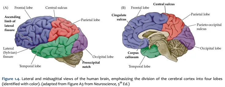
 Key areas by cytoarchitecture.
