Microscopic Anatomy of Nervous Tissues Indian Medical PG Practice Questions and MCQs
Practice Indian Medical PG questions for Microscopic Anatomy of Nervous Tissues. These multiple choice questions (MCQs) cover important concepts and help you prepare for your exams.
Microscopic Anatomy of Nervous Tissues Indian Medical PG Question 1: What are Gitter cells?
- A. Microglia
- B. Modified macrophages in the CNS (Correct Answer)
- C. Astrocytic cells
- D. Oligodendrocytic cells
Microscopic Anatomy of Nervous Tissues Explanation: ***Modified macrophages in CNS***
- Gitter cells are **modified macrophages** that have phagocytized lipid and other debris in the central nervous system (CNS), particularly in response to injury or disease [1][2].
- They play a crucial role in **cleaning up cellular debris** and are involved in the inflammatory response within the CNS [2].
*Macroglia*
- Macroglia refers to **supportive cells** in the CNS, including astrocytes and oligodendrocytes, rather than being specifically modified macrophages.
- It does not specifically describe the **phagocytic role** characteristic of Gitter cells.
*Oligodendrocytes*
- Oligodendrocytes primarily function to **myelinate axons** in the CNS and do not possess the same phagocytic capabilities as Gitter cells.
- They are involved in **insulation** of neuronal axons rather than debris clearance.
*Astrocytes*
- Astrocytes are the principal **supportive glial cells** in the CNS and do not exhibit the characteristics of Gitter cells.
- Their functions include **maintaining blood-brain barrier**, regulating blood flow, and supporting neuronal metabolism, not phagocytosis.
**References:**
[1] Kumar V, Abbas AK, et al.. Robbins and Cotran Pathologic Basis of Disease. 9th ed. Peripheral Nerves and Skeletal Muscles, pp. 1255-1256.
[2] Cross SS. Underwood's Pathology: A Clinical Approach. 6th ed. Common Clinical Manifestations Of Central And Peripheral Nervous System Disease, pp. 697-698.
Microscopic Anatomy of Nervous Tissues Indian Medical PG Question 2: Perivascular lymphocytes & microglial nodules are seen in -
- A. HIV encephalitis (Correct Answer)
- B. CMV meningitis
- C. Bacterial meningitis
- D. Multiple sclerosis
Microscopic Anatomy of Nervous Tissues Explanation: ***HIV encephalitis***
- **Perivascular lymphocytes** and **microglial nodules** are the characteristic histopathological hallmarks of **HIV encephalitis (HIV-associated dementia complex)** [1][2].
- Microglial nodules are formed by activated microglia and macrophages, often accompanied by **multinucleated giant cells** (the classic triad) [2].
- These features reflect chronic CNS inflammation and neuronal damage caused by HIV infection.
*CMV meningitis*
- Cytomegalovirus (CMV) infection in immunocompromised patients causes meningoencephalitis with characteristic **intranuclear ("owl's eye") inclusion bodies** and necrotizing inflammation.
- The histological pattern differs from the microglial nodules and perivascular lymphocytes seen in HIV encephalitis.
*Bacterial meningitis*
- Characterized by prominent **neutrophilic infiltrate** in the subarachnoid space, fibrinopurulent exudate, and potential vasculitis.
- Acute bacterial meningitis does not show the lymphocytic and microglial nodular pattern characteristic of viral encephalitis.
*Multiple sclerosis*
- An autoimmune demyelinating disease with **perivenular demyelinating plaques** containing lymphocytes and macrophages.
- While perivascular inflammation occurs, **microglial nodules** are not a characteristic feature; instead, MS shows demyelination with reactive gliosis.
**References:**
[1] Kumar V, Abbas AK, et al.. Robbins and Cotran Pathologic Basis of Disease. 9th ed. The Central Nervous System, p. 1278.
[2] Cross SS. Underwood's Pathology: A Clinical Approach. 6th ed. Common Clinical Manifestations Of Central And Peripheral Nervous System Disease, pp. 711-712.
Microscopic Anatomy of Nervous Tissues Indian Medical PG Question 3: In large neurons the nucleus can be found a large distance away from the terminal end of its axon. The body has a complex system of intracellular transporters that are able to carry essential proteins from the nucleus to the distal edge of the cell and back. Which of the following proteins are essential for this function?
- A. Kinesin, Troponin
- B. Myosin, Kinesin
- C. Actin, Dynein
- D. Dynein, Kinesin (Correct Answer)
- E. Glucose, Actin
Microscopic Anatomy of Nervous Tissues Explanation: ***Dynein, Kinesin***
- **Kinesin** is primarily responsible for **anterograde transport** (from the cell body to the axon terminal) along microtubules, carrying vesicles and organelles.
- **Dynein** handles **retrograde transport** (from the axon terminal back to the cell body), essential for recycling components and signaling.
*Kinesin, Troponin*
- While **Kinesin** is involved in axonal transport, **Troponin** is a protein found in muscle tissue that regulates muscle contraction, not intracellular transport in neurons.
- Troponin binds **calcium ions** and influences the interaction between actin and myosin.
*Myosin, Kinesin*
- **Kinesin** is involved in microtubule-based transport, but **Myosin** is primarily associated with **actin filaments** for muscle contraction and intracellular movement, not long-distance axonal transport.
- Myosin functions as a **motor protein** that converts chemical energy in ATP into mechanical force.
*Actin, Dynein*
- **Dynein** is crucial for retrograde axonal transport, but **Actin** is a structural protein forming microfilaments that are involved in cell shape, motility, and some short-distance transport, not the major long-distance axonal transport mechanism.
- Actin filaments serve as tracks for **myosin motors**, primarily in the cell cortex.
*Glucose, Actin*
- **Glucose** is a sugar molecule, the primary energy source for cells, and not a transport protein.
- **Actin** forms microfilaments for cell structure and short-range movement, not long-distance axonal transport as described.
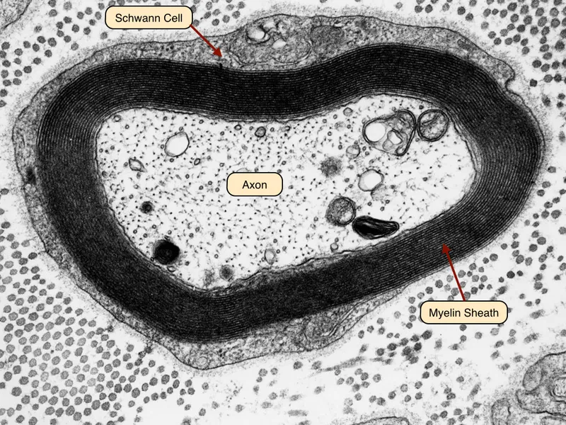
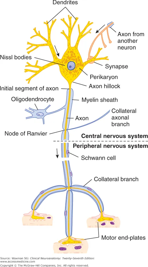
Microscopic Anatomy of Nervous Tissues Indian Medical PG Question 4: What is the primary function of Schwann cells?
- A. Form myelin sheath (Correct Answer)
- B. Part of the central nervous system
- C. Derived from neural crest cells
- D. Present in both myelinated and unmyelinated nerve fibers
Microscopic Anatomy of Nervous Tissues Explanation: ***Form myelin sheath***
- **Schwann cells** are glial cells found in the **peripheral nervous system** that wrap around axons to form the myelin sheath [1], [3].
- The **myelin sheath** acts as an electrical insulator, increasing the speed of nerve impulse conduction via **saltatory conduction** [2].
- This is the **primary and most characteristic function** of Schwann cells in the PNS [3].
*Part of the central nervous system*
- Schwann cells are exclusively found in the **peripheral nervous system (PNS)**, not the CNS [4].
- In the **central nervous system (CNS)**, **oligodendrocytes** are responsible for myelin formation [1], [4].
*Derived from neural crest cells*
- While Schwann cells are indeed derived from **neural crest cells**, this describes their **embryological origin**, not their function.
- Many other cell types (melanocytes, neurons of peripheral ganglia) are also neural crest derivatives.
*Present in both myelinated and unmyelinated nerve fibers*
- While Schwann cells are associated with both fiber types, this describes their **distribution**, not their primary function [1], [3].
- In unmyelinated fibers, Schwann cells envelop multiple axons without forming concentric myelin layers [1].
Microscopic Anatomy of Nervous Tissues Indian Medical PG Question 5: All the following features are seen in neurons from dorsal root ganglia, EXCEPT:
- A. They are multipolar (Correct Answer)
- B. They are derived from neural crest cells
- C. They have eccentrically located nuclei
- D. They contain lipofuscin granules
Microscopic Anatomy of Nervous Tissues Explanation: ***They are multipolar***
- Dorsal root ganglia (DRG) neurons are typically **pseudounipolar**, meaning they have a single process that branches into two (peripheral and central) rather than multiple dendrites and an axon [1].
- **Multipolar neurons** are characteristic of motor neurons and interneurons in the central nervous system, not DRG sensory neurons [1].
*They contain lipofuscin granules*
- **Lipofuscin granules** are common in long-lived, post-mitotic cells like neurons and are considered "wear and tear" pigments, accumulating with age.
- Their presence in DRG neurons is a normal finding and reflects the neuron's metabolic activity over time.
*They have eccentrically located nuclei*
- While not universally present in all DRG neurons, an **eccentrically located nucleus** is a common histological feature of certain types of DRG neurons, particularly larger ones.
- This feature helps distinguish them from other neuron types and can be accentuated by the large amount of cytoplasm in these cells.
*They are derived from neural crest cells*
- All sensory neurons of the DRG, along with other components like Schwann cells and sympathetic ganglia, originate from **neural crest cells**.
- This developmental origin is a fundamental characteristic of DRG neurons, distinguishing them from CNS neurons derived from the neural tube.
Microscopic Anatomy of Nervous Tissues Indian Medical PG Question 6: Which of the following is a common pathology that increases the risk of uterine injury during abdominal hysterectomy?
- A. Hydrosalpinx
- B. Pelvic endometriosis (Correct Answer)
- C. Ovarian teratoma
- D. Adenomyosis
Microscopic Anatomy of Nervous Tissues Explanation: ***Pelvic endometriosis***
- Pelvic endometriosis causes **dense adhesions, anatomical distortion, and obliteration of normal tissue planes**, making surgical dissection technically challenging during hysterectomy.
- The **fibrotic adhesions** bind pelvic organs together, obscure surgical landmarks, and increase the risk of inadvertent injury to the uterus, bladder, ureters, and bowel.
- Studies show that **endometriosis is a significant risk factor** for intraoperative complications, including uterine perforation and vascular injury.
- The **distorted pelvic anatomy** requires careful dissection and may necessitate modifications in surgical technique.
*Hydrosalpinx*
- Hydrosalpinx is a **fluid-filled, dilated fallopian tube** resulting from distal tubal obstruction, typically from prior pelvic inflammatory disease.
- While it may be encountered during hysterectomy, it does **not distort the uterine anatomy or create adhesions** that would increase the risk of uterine injury.
- Hydrosalpinx is generally easily separated from surrounding structures.
*Ovarian teratoma*
- Ovarian teratoma (dermoid cyst) is a **benign germ cell tumor of the ovary** containing mature tissues from all three germ layers.
- It is typically **well-encapsulated and does not cause significant pelvic adhesions** unless there has been rupture or torsion.
- It does not increase the risk of uterine injury during hysterectomy.
*Adenomyosis*
- Adenomyosis is **endometrial tissue within the myometrium**, causing an enlarged, boggy, tender uterus.
- While adenomyosis is often an **indication for hysterectomy**, it is an intrinsic uterine condition that does **not cause pelvic adhesions or anatomical distortion**.
- The uterus may be more vascular and bulky, but this does not specifically increase the risk of uterine injury during standard hysterectomy technique.
Microscopic Anatomy of Nervous Tissues Indian Medical PG Question 7: Feature of microscopic polyangitis is:
- A. IgG deposits in kidney
- B. Bronchospasm
- C. Renal involvement in 80% of cases (Correct Answer)
- D. All of the above
Microscopic Anatomy of Nervous Tissues Explanation: ***Renal involvement in 80% of cases***
- **Microscopic polyangiitis (MPA)** is a small-vessel vasculitis characterized by **glomerulonephritis**, which affects the kidneys in a vast majority of patients, often leading to **rapidly progressive glomerulonephritis** [1].
- The renal involvement usually presents as **pauci-immune glomerulonephritis**, meaning there are few or no immune deposits found on renal biopsy.
*IgG deposits in kidney*
- **IgG deposits** in the kidney are typically seen in immune complex-mediated glomerulonephritis, such as **lupus nephritis** or **IgA nephropathy**, not in MPA [2].
- MPA is a **pauci-immune vasculitis**, meaning there is little to no immune complex deposition in affected tissues, including the kidneys [1].
*Bronchospasm*
- **Bronchospasm** is primarily a feature of **asthma** or other obstructive airway diseases, and not a direct or prominent feature of microscopic polyangiitis.
- While lung involvement (e.g., **pulmonary hemorrhage**) can occur in MPA, it typically manifests as dyspnea, cough, or hemoptysis, rather than bronchospasm [1].
*All of the above*
- This option is incorrect because neither **IgG deposits in the kidney** nor **bronchospasm** are characteristic features of microscopic polyangiitis.
- The presence of **renal involvement in 80% of cases** is an accurate characteristic, but it is not the only option presented and "all of the above" implies the other features are also true.
Microscopic Anatomy of Nervous Tissues Indian Medical PG Question 8: Patient presenting with cutaneous vasculitis, glomerulonephritis, peripheral neuropathy, Which investigation is to be performed next that will help you diagnose the condition?
- A. ANCA (Correct Answer)
- B. RA factor
- C. Hbsag
- D. MIF
Microscopic Anatomy of Nervous Tissues Explanation: ### ANCA
- The combination of **cutaneous vasculitis**, **glomerulonephritis**, and **peripheral neuropathy** points towards a small-vessel vasculitis, for which **ANCA (anti-neutrophil cytoplasmic antibodies)** testing is crucial [1].
- ANCA is highly specific for conditions like **Granulomatosis with Polyangiitis (GPA)** and **Microscopic Polyangiitis (MPA)** [1].
### RA factor
- **Rheumatoid factor (RF)** is primarily associated with **rheumatoid arthritis**, which typically presents with symmetrical polyarthritis, not the constellation of symptoms described.
- While RF can be positive in some vasculitides, it is not the most specific initial test for the given clinical presentation.
### Hbsag
- **Hepatitis B surface antigen (HbsAg)** typically screens for **Hepatitis B infection**, which can cause **polyarteritis nodosa (PAN)**, a medium-vessel vasculitis.
- However, the patient's symptoms (cutaneous vasculitis, glomerulonephritis) are more characteristic of **small-vessel vasculitis**, making ANCA a more direct investigation [1].
### MIF
- **MIF (Macrophage Migration Inhibitory Factor)** is a cytokine involved in inflammation, but it is not a routine diagnostic marker for vasculitis.
- It is not used as a primary investigation to diagnose specific autoimmune or inflammatory conditions like vasculitis.
Microscopic Anatomy of Nervous Tissues Indian Medical PG Question 9: A patient presents with pulmonary hemorrhage and is P-ANCA positive. What is the most likely diagnosis?
- A. Churg-Strauss syndrome
- B. Microscopic polyangiitis (Correct Answer)
- C. Wegener granulomatosis
- D. Polyarteritis nodosa (PAN)
Microscopic Anatomy of Nervous Tissues Explanation: ***Microscopic polyangiitis***
- This condition is characterized by **pulmonary hemorrhage** (often manifesting as diffuse alveolar hemorrhage) and **P-ANCA positivity**, which is typically associated with antibodies against **myeloperoxidase (MPO)**. [1]
- It is a **small-vessel vasculitis** that frequently affects the kidneys (glomerulonephritis) and lungs without granuloma formation.
*Churg-Strauss syndrome*
- While Churg-Strauss syndrome (now known as **Eosinophilic Granulomatosis with Polyangiitis**, EGPA) can be P-ANCA positive, it is typically associated with a history of **asthma**, **allergic rhinitis**, and **eosinophilia**. [1]
- Pulmonary involvement often includes **infiltrates** and nodules, but diffuse alveolar hemorrhage with severe pulmonary hemorrhage is less common as the primary presentation compared to MPA.
*Wegener granulomatosis*
- Wegener granulomatosis (now known as **Granulomatosis with Polyangiitis**, GPA) primarily presents with **upper and lower respiratory tract granulomatous inflammation** and **glomerulonephritis**.
- It is typically associated with **C-ANCA positivity** (antibodies against proteinase 3, PR3), not P-ANCA.
*Polyarteritis nodosa (PAN)*
- Polyarteritis nodosa is a **medium-vessel vasculitis** that typically affects the **kidneys, gastrointestinal tract, skin, and nervous system**. [1]
- It is classically **ANCA-negative** and does not typically cause pulmonary hemorrhage or diffuse alveolar hemorrhage.
Microscopic Anatomy of Nervous Tissues Indian Medical PG Question 10: Which of the following cell types is neuroectodermal in origin?
- A. Smooth muscle cells (Correct Answer)
- B. Skeletal muscle cells
- C. Endothelial cells
- D. Cardiac muscle cells
Microscopic Anatomy of Nervous Tissues Explanation: ***Smooth muscle cells***
- This is the **correct answer** based on a **specific exception**: smooth muscle cells of the **iris dilator and sphincter muscles** and the **ciliary muscle** in the eye are derived from **neuroectoderm** (specifically from the **optic cup**, an outgrowth of the neural tube).
- **Important note:** The vast majority of smooth muscle in the body is of **mesodermal origin** (e.g., in blood vessels, GI tract, respiratory tract). This question tests knowledge of this **notable embryological exception**.
- In the context of the given options, this is the only cell type with any neuroectodermal component.
*Skeletal muscle cells*
- Skeletal muscle cells are entirely derived from the **paraxial mesoderm**, specifically from **somites** (myotome portion).
- They form the voluntary muscles of the body and are **never** of neuroectodermal origin.
*Endothelial cells*
- Endothelial cells lining blood vessels and lymphatic vessels are derived from the **mesoderm** (specifically from **angioblasts**).
- They are part of the cardiovascular system and are **entirely mesodermal** in origin.
*Cardiac muscle cells*
- Cardiac muscle cells are derived from the **splanchnic mesoderm** (lateral plate mesoderm).
- The heart musculature is **entirely mesodermal** with no neuroectodermal contribution.
**Clinical Pearl:** Classic neuroectodermal derivatives include neurons, glial cells (astrocytes, oligodendrocytes), ependymal cells, and neural crest derivatives (Schwann cells, melanocytes, chromaffin cells). The smooth muscle of the iris represents an important exception to the general rule that smooth muscle is mesodermal.
More Microscopic Anatomy of Nervous Tissues Indian Medical PG questions available in the OnCourse app. Practice MCQs, flashcards, and get detailed explanations.


 , oligodendrocytes (fried-egg), microglia (small, elongated), ependymal cells (cuboidal, ciliated))