Joints of Lower Limb Indian Medical PG Practice Questions and MCQs
Practice Indian Medical PG questions for Joints of Lower Limb. These multiple choice questions (MCQs) cover important concepts and help you prepare for your exams.
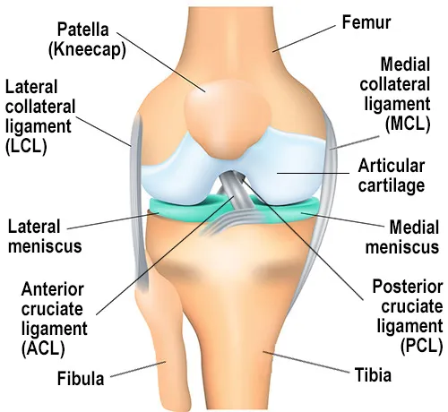
Joints of Lower Limb Indian Medical PG Question 1: Which ligament is most commonly damaged in knee injuries?
- A. PCL
- B. LCL
- C. MCL
- D. ACL (Correct Answer)
Joints of Lower Limb Explanation: ***ACL***
- The **anterior cruciate ligament (ACL)** is highly susceptible to injury, especially during sports involving sudden stops, changes in direction, jumping, and awkward landings.
- Its role in stabilizing the knee against **anterior tibial translation** and rotational forces makes it vulnerable to tears.
*PCL*
- The **posterior cruciate ligament (PCL)** is much stronger than the ACL and less frequently injured, typically requiring a direct blow to the flexed knee (e.g., dashboard injury).
- It prevents **posterior tibial translation** relative to the femur.
*MCL*
- The **medial collateral ligament (MCL)** is commonly injured, often due to a direct blow to the outside of the knee causing a **valgus stress**.
- While frequently damaged, it is often injured in conjunction with the ACL but the ACL is more frequently injured in isolation.
*LCL*
- The **lateral collateral ligament (LCL)** is the least commonly injured of the four major knee ligaments.
- It usually results from a direct blow to the inside of the knee causing **varus stress**.
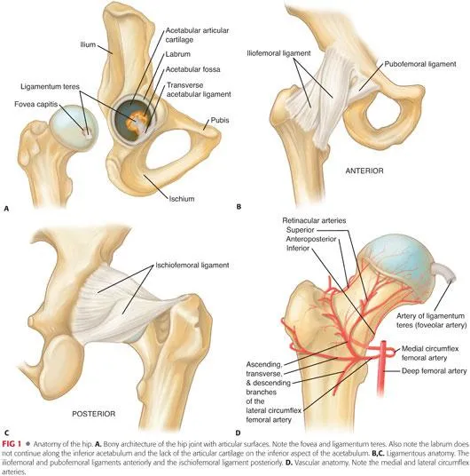
Joints of Lower Limb Indian Medical PG Question 2: The arteries supplying the femoral head include the following except
- A. Ligamentum teres artery (Correct Answer)
- B. Medial circumflex artery
- C. Profunda femoral artery
- D. Lateral circumflex artery
Joints of Lower Limb Explanation: ***Ligamentum teres artery***
- While called an artery, the artery of the **ligamentum teres** (foveal artery) is an **inconsistent** and typically **insignificant** contributor to the femoral head blood supply in adults.
- Its primary role, when present, is mainly during **development** and it often **obliterates** or remains a small vessel that usually provides **minimal to no significant blood supply** to the femoral head in adults.
- Of all the arteries listed, this is the **least reliable** and most frequently absent or non-functional supplier.
*Medial circumflex femoral artery*
- The **medial circumflex femoral artery** is the **most crucial** blood supply to the adult femoral head, providing approximately **75-80%** of the blood supply, especially to the superior and posterior aspects.
- It gives rise to the **retinacular arteries** that ascend along the femoral neck beneath the synovial reflection.
*Profunda femoris artery*
- The **profunda femoris artery** (deep femoral artery) is the main branch of the femoral artery and gives rise to the **medial and lateral circumflex femoral arteries**.
- While it is the **parent vessel** of the actual suppliers, it does not **directly** supply the femoral head itself—its branches do.
- In strict anatomical terms, it is a **source artery** rather than a direct supplier, but it is included here as it gives rise to the circumflex vessels.
*Lateral circumflex femoral artery*
- The **lateral circumflex femoral artery** also contributes to the blood supply of the femoral head, though typically to a **lesser extent** than the medial circumflex femoral artery.
- It supplies the **anterior aspect** of the femoral head and neck, primarily through its ascending branch.
Joints of Lower Limb Indian Medical PG Question 3: What is the condition commonly known as jumper's knee?
- A. Inflammation of the patellar tendon at its insertion on the patella.
- B. Tendinopathy of the quadriceps tendon.
- C. Injury to the hamstring tendon.
- D. Patellar tendonitis due to overuse of the patellar tendon. (Correct Answer)
Joints of Lower Limb Explanation: ***Patellar tendonitis due to overuse of the patellar tendon.***
- **Jumper's knee** is the common term for **patellar tendonitis**, which specifically refers to inflammation of the patellar tendon.
- This condition is frequently caused by **overuse**, especially in activities involving repetitive jumping and landing.
*Inflammation of the patellar tendon at its insertion on the patella.*
- While jumper's knee does involve inflammation of the patellar tendon, it is more commonly at its insertion on the **tibial tubercle** or specifically its origin at the **inferior pole of the patella**, not necessarily at the patella itself.
- This option is less precise as it describes only one aspect of the condition without mentioning the critical role of overuse.
*Tendinopathy of the quadriceps tendon.*
- **Tendinopathy of the quadriceps tendon** is a distinct condition affecting the tendon above the patella, known as **quadriceps tendinopathy**.
- It presents with pain proximal to the patella, differentiating it from jumper's knee, which involves the tendon distal to the patella.
*Injury to the hamstring tendon.*
- An **injury to the hamstring tendon** would cause pain and symptoms on the posterior aspect of the knee or thigh.
- This is completely unrelated to jumper's knee, which is characterized by anterior knee pain.
Joints of Lower Limb Indian Medical PG Question 4: Which of the following ligaments is injured in an ankle inversion injury?
- A. Calcaneofibular ligament
- B. Posterior talofibular ligament
- C. Deltoid ligament
- D. Anterior talofibular ligament (Correct Answer)
Joints of Lower Limb Explanation: ***Anterior talofibular ligament***
- The **anterior talofibular ligament (ATFL)** is the most commonly injured ligament in an **ankle inversion sprain** due to its position and weaker structure.
- It connects the **fibula** to the **talus** anteriorly, and when the foot inverts, this ligament is stretched and often torn first.
*Calcaneofibular ligament*
- The **calcaneofibular ligament (CFL)** is also an important lateral ankle ligament that can be injured in **severe inversion sprains**.
- It is often damaged in conjunction with the ATFL, but typically only after the ATFL has already been compromised through an ankle inversion injury.
*Posterior talofibular ligament*
- The **posterior talofibular ligament (PTFL)** is the strongest of the **lateral collateral ligaments** and is rarely injured in isolation.
- Injury to the PTFL usually occurs in cases of **severe, high-grade ankle dislocations** or very forceful inversion injuries, often involving other ligaments.
*Deltoid ligament*
- The **deltoid ligament** is a strong, fan-shaped ligament located on the **medial side of the ankle**.
- It resists **eversion** of the ankle, meaning it is more commonly injured in **eversion sprains**, not inversion sprains.
Joints of Lower Limb Indian Medical PG Question 5: All are true about osteoarthritis, except
- A. Quadriceps atrophy (Correct Answer)
- B. MCP is spared
- C. Glucosamines are beneficial
- D. Loose bodies in the ankle joint
Joints of Lower Limb Explanation: ***Quadriceps atrophy***
- While muscle weakness can occur in osteoarthritis due to pain and disuse, **quadriceps atrophy** is not a universal or defining characteristic of the disease itself, nor is it consistently observed as a primary feature.
- The statement implies that quadriceps atrophy is *always* true about osteoarthritis, which is incorrect as it's a potential consequence but not inherently present in all cases or a direct pathological feature.
*MCP is spared*
- The **metacarpophalangeal (MCP) joints** are typically spared in osteoarthritis, unlike in rheumatoid arthritis.
- Osteoarthritis predominantly affects the **distal interphalangeal (DIP)** and **proximal interphalangeal (PIP)** joints of the hands, as well as the **carpometacarpal (CMC) joint of the thumb**.
*Glucosamines are beneficial*
- **Glucosamine sulfate** is a commonly used supplement in osteoarthritis, with some studies suggesting it may provide modest pain relief and slow cartilage degradation in certain patients.
- While its efficacy is debated and not universally accepted as curative, many patients report subjective benefit, and it is considered a complementary therapy.
*Loose bodies in the ankle joint*
- **Loose bodies**, also known as joint mice, are fragments of cartilage or bone that can break off and float within the joint space.
- These are a recognized complication of osteoarthritis, particularly in weight-bearing joints like the **ankle**, and can cause locking or catching sensations.
Joints of Lower Limb Indian Medical PG Question 6: A football player experienced a twist in the ankle and knee. Clinically, no bone injury was appreciated. The examiner is performing the test shown in the image. Which test is this?
- A. Posterior drawer for PCL
- B. McMurray
- C. Lachman (Correct Answer)
- D. Anterior drawer for ACL
Joints of Lower Limb Explanation: ***Lachman***
- The image shows the examiner holding the distal thigh and proximal tibia, with the knee flexed at a **20-30 degree angle**, applying an **anterior translational force** to the tibia. This specific maneuver is characteristic of the Lachman test.
- The Lachman test is highly sensitive for detecting **anterior cruciate ligament (ACL) tears**, particularly in acute injuries, due to the reduced hamstring spasm compared to the anterior drawer test.
*Posterior drawer for PCL*
- The posterior drawer test involves flexing the knee to **90 degrees** and applying a **posterior force** to the tibia to assess the integrity of the **posterior cruciate ligament (PCL)**.
- The position of the knee in the image (flexed at a shallower angle) and the direction of the applied force (anteriorly towards the femur) do not match the technique for a posterior drawer test.
*McMurray*
- The McMurray test is performed to evaluate **meniscal tears** by flexing, extending, and rotating the knee while applying a varus or valgus stress.
- The maneuver in the image, involving direct anterior translation of the tibia with the knee in slight flexion, is not consistent with the McMurray test.
*Anterior drawer for ACL*
- While also testing the **ACL**, the anterior drawer test typically involves flexing the knee to **90 degrees** and sitting on the foot, then pulling the tibia anteriorly.
- The knee flexion angle in the image is much shallower than 90 degrees, making it inconsistent with the standard anterior drawer test.
Joints of Lower Limb Indian Medical PG Question 7: Which of the following group of lymph nodes does NOT receive direct lymphatic drainage from the perineum?
- A. Superficial inguinal
- B. Internal iliac
- C. External iliac (Correct Answer)
- D. Deep inguinal
Joints of Lower Limb Explanation: ***External iliac***
- The external iliac lymph nodes do **NOT receive direct lymphatic drainage** from the perineum.
- They primarily receive lymph from the **deep inguinal nodes**, pelvic organs (bladder, upper vagina), and lower anterior abdominal wall [1].
- Perineal lymphatics drain to superficial inguinal, deep inguinal, or internal iliac nodes first, making external iliac a **secondary or tertiary drainage station** rather than a direct recipient.
*Superficial inguinal*
- These are the **primary drainage site** for lymph from the superficial perineum.
- They receive direct lymphatic vessels from the **vulva, distal vagina, labia majora**, scrotum, and skin of the perineum.
- This is the main first-line drainage pathway for superficial perineal structures.
*Internal iliac*
- Internal iliac lymph nodes receive **direct lymphatic drainage** from the deep perineum, including the **male urethra, prostate**, and deep structures [2], [3].
- They serve as primary drainage for pelvic visceral structures and deep perineal tissues [3].
*Deep inguinal*
- Deep inguinal lymph nodes receive lymph from the **superficial inguinal nodes** and from deep structures of the lower limb.
- They are part of the drainage pathway from the perineum via the superficial inguinal nodes.
Joints of Lower Limb Indian Medical PG Question 8: Which structure lies midway between the anterior superior iliac spine and pubic symphysis?
- A. Femoral artery (Correct Answer)
- B. Deep inguinal ring
- C. Superior epigastric artery
- D. Inguinal ligament
Joints of Lower Limb Explanation: ***Femoral artery***
- The **femoral artery** is a direct continuation of the external iliac artery and is the most reliable palpable pulse in the groin area. [1]
- Its surface marking is clinically important as it's found midway between the **anterior superior iliac spine (ASIS)** and the **pubic symphysis**, specifically at the **mid-inguinal point**. [1]
*Deep inguinal ring*
- The **deep inguinal ring** is located at the **midpoint of the inguinal ligament** (midway between ASIS and pubic tubercle), which is approximately 1.5 cm above and lateral to the mid-inguinal point.
- It marks the beginning of the **inguinal canal** and is the site where the vas deferens and gonadal vessels exit the abdominal cavity.
*Superior epigastric artery*
- The **superior epigastric artery** is a terminal branch of the internal thoracic artery and primarily supplies the upper abdominal wall. [2]
- It is located in the anterior abdominal wall, far from the inguinal region and the midpoint between the ASIS and pubic symphysis. [2]
*Inguinal ligament*
- The **inguinal ligament** extends between the anterior superior iliac spine and the pubic tubercle, forming the inferior border of the anterior abdominal wall.
- While relevant to the region, the ligament itself is a fibrous band, not a structure found *midway between* the ASIS and pubic symphysis in the same way the femoral artery is.
Joints of Lower Limb Indian Medical PG Question 9: All are predisposing factors of Deep Vein thrombosis, EXCEPT :
- A. Lower limb trauma
- B. Cushing's syndrome
- C. Hip surgery
- D. Subungual melanoma (Correct Answer)
Joints of Lower Limb Explanation: ***Subungual melanoma***
- This is a rare form of melanoma that develops under the nail, and while serious, it is **not a recognized predisposing factor for deep vein thrombosis (DVT)**. Its primary concerns are local invasion and metastasis.
- Unlike conditions affecting blood clotting or endothelium, **subungual melanoma does not directly promote hypercoagulability, venous stasis, or endothelial damage** that contribute to DVT.
*Lower limb trauma*
- **Trauma to the lower limb** can cause **endothelial damage** to blood vessels and **venous stasis** due to immobility or swelling, both key components of **Virchow's triad** for DVT [1].
- **Fractures or severe soft tissue injuries** often necessitate immobilization and can lead to inflammation, further increasing the risk of clot formation [1].
*Cushing's syndrome*
- **Cushing's syndrome** is associated with **hypercoagulability** due to increased levels of clotting factors, such as **factor VIII** and **fibrinogen**, and decreased fibrinolytic activity.
- The **elevated cortisol levels** seen in Cushing's syndrome [2] can directly contribute to a prothrombotic state, significantly increasing DVT risk.
*Hip surgery*
- **Major orthopedic surgeries**, especially hip surgery [1], are well-known to cause significant **venous stasis** and **endothelial damage**.
- **Post-operative immobility** and a generalized **inflammatory response** following surgery contribute to a high risk of DVT formation [1].
Joints of Lower Limb Indian Medical PG Question 10: Which one of the following is a manifestation of a "negative G"?
- A. The hydrostatic pressure in veins of lower limb increases
- B. The cardiac output decreases
- C. Black out occurs
- D. The cerebral arterial pressure rises (Correct Answer)
Joints of Lower Limb Explanation: ***The cerebral arterial pressure rises***
- A **negative G-force** pushes blood towards the head, causing an increase in hydrostatic pressure in the cerebral arteries.
- This **elevated pressure** can lead to symptoms such as facial swelling, headache, and even petechial hemorrhages in severe cases.
*The hydrostatic pressure in veins of lower limb increases*
- **Negative G-forces** push blood away from the lower limbs towards the head, thereby **decreasing** hydrostatic pressure in the lower limb veins.
- This is in contrast to positive G-forces, which increase hydrostatic pressure in the lower limbs.
*The cardiac output decreases*
- While extreme gravitational forces (both positive and negative) can impact cardiac output, a direct and common manifestation of **negative G** is not a decrease in cardiac output.
- The initial effect of negative G is often increased venous return to the heart from regions below the heart, which might transiently *increase* cardiac output.
*Black out occurs*
- **Blackout (loss of vision followed by loss of consciousness)** is a characteristic symptom of **positive G-forces**, where blood is pulled away from the head, leading to cerebral ischemia.
- In contrast, **redout** (reddening of vision due to conjunctival congestion) can occur with severe negative G-forces due to increased cerebral blood pressure.
More Joints of Lower Limb Indian Medical PG questions available in the OnCourse app. Practice MCQs, flashcards, and get detailed explanations.


oka
 , inferior tibiofibular, tarsometatarsal (Lisfranc's), metatarsophalangeal, and interphalangeal joints)