Surgical Anatomy Indian Medical PG Practice Questions and MCQs
Practice Indian Medical PG questions for Surgical Anatomy. These multiple choice questions (MCQs) cover important concepts and help you prepare for your exams.
Surgical Anatomy Indian Medical PG Question 1: Not a landmark of facial nerve identification in parotid surgery:
- A. Peripheral branches
- B. Post belly of digastric
- C. Inferior belly of omohyoid (Correct Answer)
- D. Tragal pointer
Surgical Anatomy Explanation: ***Inferior belly of omohyoid***
- The **inferior belly of the omohyoid muscle** is located in the anterior triangle of the neck and is not a surgical landmark for the facial nerve during parotidectomy.
- Its anatomical position is too far inferior and anterior to the parotid gland and facial nerve trunk to be useful for facial nerve identification.
*Peripheral branches*
- While careful dissection of **peripheral branches** is crucial for preserving facial nerve function, they are typically identified *after* locating the main trunk, not as primary landmarks for initially finding the nerve.
- Direct identification of peripheral branches first is challenging and carries a higher risk of injury without prior identification of the main trunk or its primary divisions.
*Post belly of digastric*
- The **posterior belly of the digastric muscle** serves as a vital deep landmark for locating the facial nerve trunk.
- The facial nerve typically passes superior to and deep to the posterior belly of the digastric muscle, providing a reliable point of reference for approaching the nerve.
*Tragal pointer*
- The **tragal pointer**, referring to the anterior surface of the cartilaginous tragus, is a superficial landmark used to approximate the location of the facial nerve trunk.
- The facial nerve's main trunk typically emerges from the stylomastoid foramen, which is positioned anterior and inferior to the tragus, making it a useful starting point for surgical dissection.
Surgical Anatomy Indian Medical PG Question 2: The lower border of the pharynx is the level of:
- A. C2
- B. C3
- C. C4
- D. C6 (Correct Answer)
Surgical Anatomy Explanation: ***C6***
- The **pharynx** extends from the base of the skull to the inferior border of the **cricoid cartilage** [1].
- This anatomical landmark, the inferior border of the **cricoid cartilage**, is located at the level of the **C6 vertebra** [1].
*C2*
- The C2 vertebra, also known as the **axis**, is significantly higher than the lower border of the pharynx.
- It is involved in head rotation and forms part of the **atlantoaxial joint**.
*C3*
- The C3 vertebra is located higher in the cervical spine and is associated with structures like the hyoid bone, but not the lower pharyngeal border.
- It is the approximate level of the **hyoid bone** [1].
*C4*
- The C4 vertebra is typically at the level of the superior border of the **thyroid cartilage**, which is still superior to the lower pharynx.
- This level is also associated with the bifurcation of the common carotid artery.
Surgical Anatomy Indian Medical PG Question 3: For a midline incision in the abdomen, length of suture required is:
- A. 3 times the length of incision
- B. 4 times the length of incision (Correct Answer)
- C. 2 times the length of incision
- D. 5 times the length of incision
Surgical Anatomy Explanation: ***4 times the length of incision***
- The standard recommendation for interrupted abdominal fascial closure is to use a **suture-to-wound length ratio** of approximately **4:1**.
- This ratio ensures sufficient material for adequate fascial apposition, overlapping bites, and knots, which are crucial for preventing wound dehiscence.
*3 times the length of incision*
- A 3:1 suture-to-wound ratio might be insufficient for secure fascial closure, potentially leading to increased tension on the suture lines and a **higher risk of dehiscence**.
- This ratio could be considered for very specific continuous closure techniques, but it's generally not recommended for standard interrupted closures.
*2 times the length of incision*
- A 2:1 ratio is generally considered **inadequate** for most fascial closures, especially in the abdomen.
- This ratio would likely result in insufficient suture material, leading to very large bites and an insecure closure, significantly increasing the risk of **wound dehiscence** and **herniation**.
*5 times the length of incision*
- While it ensures enough material, a 5:1 ratio suggests using **excessive suture material** which might extend operating time.
- Using significantly more suture than necessary offers no proven benefit in terms of wound security and can sometimes introduce more foreign material into the wound.
Surgical Anatomy Indian Medical PG Question 4: During a Pfannenstiel incision, which of the following nerves is most at risk of injury due to its anatomical location?
- A. T10
- B. T11
- C. Iliohypogastric (Correct Answer)
- D. Ilioinguinal
Surgical Anatomy Explanation: ***Iliohypogastric***
- The **iliohypogastric nerve** travels superior and parallel to the **inguinal ligament** and is vulnerable during a Pfannenstiel incision due to its course through the **oblique muscles** at the lateral edge of the incision [1].
- Injury can lead to **sensory loss** over the suprapubic area and motor weakness of the transected abdominal wall muscles.
*T10*
- The **T10 dermatome** covers the umbilical region, which is generally superior to the typical Pfannenstiel incision site.
- While theoretically possible, direct injury to the **T10 nerve** is less common compared to nerves coursing through the lower abdominal wall muscles.
*T11*
- The **T11 nerve** innervates the region between the umbilicus and the pubic area, but its course is typically more medial and less exposed at the lateral edges of a Pfannenstiel incision.
- Injury to **T11** is therefore less likely during this specific surgical approach compared to the iliohypogastric nerve.
*Ilioinguinal*
- The **ilioinguinal nerve** runs more inferior and medial to the **iliohypogastric nerve**, closer to the inguinal canal [1].
- While also at risk during lower abdominal incisions, the **iliohypogastric nerve** is generally considered to be at higher risk during a Pfannenstiel incision due to its more superficial and lateral course at the incision margins.
Surgical Anatomy Indian Medical PG Question 5: Surgical neck fracture leads to all EXCEPT
- A. Deltoid muscle palsy
- B. Weakness of abduction
- C. Teres minor palsy
- D. Teres major palsy (Correct Answer)
Surgical Anatomy Explanation: ***Teres major palsy***
- The **teres major** muscle is innervated by the **lower subscapular nerve** (C5-C7).
- A surgical neck fracture of the humerus typically injures the **axillary nerve**, which does not innervate the teres major.
*Deltoid muscle palsy*
- The **axillary nerve**, which innervates the **deltoid muscle**, is commonly injured in a surgical neck fracture due to its proximity.
- Injury to the axillary nerve would result in **deltoid muscle palsy**, leading to weakness in shoulder abduction and external rotation.
*Weakness of abduction*
- The **deltoid muscle** is the primary abductor of the arm after the initial 15 degrees, and it is innervated by the **axillary nerve**.
- A surgical neck fracture carries a high risk of **axillary nerve injury**, compromising deltoid function and causing significant weakness in abduction.
*Teres minor palsy*
- The **teres minor muscle** is innervated by the **axillary nerve**, which is vulnerable in surgical neck fractures.
- Palsy of the teres minor would impair **external rotation** of the shoulder.
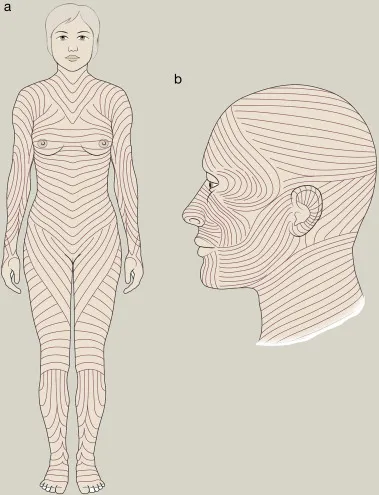
Surgical Anatomy Indian Medical PG Question 6: Identify the lines shown in the following image:
- A. Hinderer's lines
- B. Dermatomes
- C. Langer's lines (Correct Answer)
- D. Blaschko's lines
Surgical Anatomy Explanation: ***Langer's lines***
- The image displays lines that represent the **natural orientation of collagen fibers** within the human skin, which are known as Langer's lines (also called cleavage lines).
- Making surgical incisions **parallel to these lines** can result in better wound healing and less scarring.
- Named after **Karl Langer**, an Austrian anatomist who described these lines in 1861.
*Hinderer's lines*
- While **Hinderer** described relaxed skin tension lines (RSTLs) used in plastic surgery, these are **different from Langer's lines**.
- The image shows Langer's lines specifically, which are based on **collagen fiber orientation**, not relaxed skin tension.
*Dermatomes*
- **Dermatomes** are areas of skin mainly supplied by a single **spinal nerve root**.
- They represent **neurologic segments** and do not correspond to the collagen fiber orientation shown in the image.
*Blaschko's lines*
- **Blaschko's lines** are invisible lines of skin cell migration that become visible in certain **genetic or acquired dermatological conditions**.
- They represent a **mosaic pattern** due to different cell populations and are distinctly different from the structural collagen lines shown.
Surgical Anatomy Indian Medical PG Question 7: Inferior epigastric artery forms the boundary of?
- A. Femoral triangle
- B. Hesselbach's triangle (Correct Answer)
- C. Adductor canal
- D. Popliteal triangle
Surgical Anatomy Explanation: ***Hesselbach's triangle***
- The **inferior epigastric artery** forms the superolateral border of Hesselbach's triangle [1].
- This triangle is clinically significant as it is a common site for **direct inguinal hernias** due to its relative weakness [1].
*Femoral triangle*
- The femoral triangle is bounded by the **inguinal ligament superiorly**, the **sartorius muscle laterally**, and the **adductor longus muscle medially**.
- It contains the **femoral nerve**, artery, and vein.
*Adductor canal*
- The adductor canal is an intermuscular tunnel located in the **thigh**, containing the **femoral artery and vein** and the **saphenous nerve**.
- Its boundaries are the **vastus medialis**, adductor longus/magnus, and sartorius muscles.
*Popliteal triangle*
- This term is not a standard anatomical triangle. The correct term is the **popliteal fossa**, which is a diamond-shaped space behind the knee joint.
- The popliteal fossa contains structures such as the **popliteal artery and vein**, tibial nerve, and common fibular nerve.
Surgical Anatomy Indian Medical PG Question 8: A 58-year-old male with a history of hypertension and smoking presents with sudden severe back pain and hypotension. A CT scan reveals a 7 cm ruptured abdominal aortic aneurysm (AAA). What are the key factors in deciding whether to proceed with endovascular aneurysm repair (EVAR) or open surgical repair?
- A. Patient's hemodynamic stability, anatomy of the aneurysm, and access to EVAR equipment (Correct Answer)
- B. Patient's hemodynamic stability and anatomy of the aneurysm
- C. Access to EVAR equipment and patient's age
- D. Surgeon's experience with EVAR procedures
Surgical Anatomy Explanation: ***Patient's hemodynamic stability, anatomy of the aneurysm, and access to EVAR equipment***
- **Hemodynamic stability** is crucial; unstable patients may benefit from more rapid intervention, potentially open repair, or require stabilization before EVAR.
- The **anatomy of the aneurysm** (e.g., neck length, angulation, iliac artery access) dictates suitability for EVAR, as specific morphological criteria must be met for stent-graft placement.
- **Access to EVAR equipment and trained personnel** is also a practical consideration for emergency intervention.
*Patient's hemodynamic stability and anatomy of the aneurysm*
- While **hemodynamic stability** and **aneurysm anatomy** are critical factors, access to specialized EVAR equipment and facilities is also a practical determinant of whether EVAR can even be attempted, especially in an emergent setting.
- This option overlooks the logistical requirements necessary for performing an **EVAR procedure**.
*Access to EVAR equipment and patient's age*
- **Access to EVAR equipment** is important, but **patient's age** is generally less critical than factors like physiological status, comorbidities, and aneurysm morphology when deciding between EVAR and open repair for ruptured AAAs.
- Younger patients may tolerate open surgery better, but age alone does not preclude EVAR if anatomy is suitable.
*Surgeon's experience with EVAR procedures*
- While **surgeon experience** is important for procedural success and outcomes, it is considered secondary to the immediate patient-centered and anatomical factors.
- In emergency settings, the decision primarily hinges on the **patient's hemodynamic status**, **aneurysm anatomical suitability**, and **immediate availability of EVAR resources**, rather than being driven by surgeon preference based on experience alone.
- Institutional protocols typically guide whether EVAR or open repair should be attempted based on the factors in the correct answer.
Surgical Anatomy Indian Medical PG Question 9: The following arrow marked vessel can cause torrential hemorrhage during cholecystectomy. Which of the following is the correct description?
- A. Moynihan's caterpillar hump due to bend of right hepatic artery (Correct Answer)
- B. Moynihan's caterpillar hump due to bend of left hepatic artery
- C. Moynihan's caterpillar hump due to bend of cystic artery
- D. Moynihan's caterpillar hump due to bend of common hepatic artery
Surgical Anatomy Explanation: ***Moynihan's caterpillar hump due to bend of right hepatic artery***
- The image shows an anatomical variation where the **right hepatic artery** forms a tortuous bend near the cystic duct, resembling a "caterpillar hump."
- This anatomical anomaly, known as **Moynihan's hump**, places the right hepatic artery in close proximity to the operative field during cholecystectomy, making it vulnerable to accidental injury and potentially causing torrential hemorrhage.
*Moynihan's caterpillar hump due to bend of left hepatic artery*
- The left hepatic artery originates from the common hepatic artery and supplies the left lobe of the liver, typically staying well away from the area of concern during routine cholecystectomy.
- A bend in the **left hepatic artery** would not be located in the position shown or pose the same risk during gallbladder removal.
*Moynihan's caterpillar hump due to bend of cystic artery*
- The cystic artery typically arises from the right hepatic artery and is ligated during cholecystectomy to devascularize the gallbladder.
- While it supplies the gallbladder, the described "caterpillar hump" refers specifically to a tortuous **right hepatic artery**, not the cystic artery itself.
*Moynihan's caterpillar hump due to bend of common hepatic artery*
- The common hepatic artery branches into the proper hepatic artery and gastroduodenal artery, located more proximally to the area depicted.
- A bend in the **common hepatic artery** would not be found in such close proximity to the cystic duct and would not be described as Moynihan's caterpillar hump in this context.
Surgical Anatomy Indian Medical PG Question 10: Branchial arches give rise to various structures in the head and neck region. From which arch does the maxillary artery develop?
- A. 3rd arch
- B. 4th arch
- C. 1st arch (Correct Answer)
- D. 5th arch
Surgical Anatomy Explanation: ***Correct Option: 1st arch***
- The **maxillary artery** develops from the **first pharyngeal arch artery** (mandibular arch).
- The first arch artery is the arterial component of the mandibular arch and gives rise to the **maxillary artery**, which supplies the maxillofacial region.
- This is consistent with the first arch's role in forming structures of the **maxilla and mandible**.
*Incorrect Option: 3rd arch*
- The third arch artery contributes to the **common carotid artery** and the **internal carotid artery**.
- It is not involved in the formation of the maxillary artery.
*Incorrect Option: 4th arch*
- The fourth arch artery forms part of the **aortic arch** on the left and the **subclavian artery** on the right.
- Its contributions are primarily to the systemic great vessels, not the maxillofacial vasculature.
*Incorrect Option: 5th arch*
- The fifth pharyngeal arch is often **rudimentary** or **absent** in humans, and when present, it regresses entirely.
- It does not contribute to any significant adult arterial structures.
More Surgical Anatomy Indian Medical PG questions available in the OnCourse app. Practice MCQs, flashcards, and get detailed explanations.