Spinal Cord and Meninges Indian Medical PG Practice Questions and MCQs
Practice Indian Medical PG questions for Spinal Cord and Meninges. These multiple choice questions (MCQs) cover important concepts and help you prepare for your exams.
Spinal Cord and Meninges Indian Medical PG Question 1: Vertebral arteries of both sides unite to form
- A. Anterior spinal artery
- B. Posterior spinal artery
- C. Medullary artery
- D. Basilar artery (Correct Answer)
Spinal Cord and Meninges Explanation: Basilar artery
- The paired vertebral arteries ascend through the neck via the transverse foramina of cervical vertebrae and enter the skull through the foramen magnum.
- At the level of the pontomedullary junction, the two vertebral arteries merge to form a single basilar artery.
Anterior spinal artery
- The anterior spinal artery is formed by the union of two small branches derived from each vertebral artery near their intracranial origin.
- It supplies the anterior two-thirds of the spinal cord, running along the anterior median fissure.
Posterior spinal artery
- The posterior spinal arteries are typically two vessels, one arising from each vertebral artery (or less commonly from the posterior inferior cerebellar artery).
- They supply the posterior one-third of the spinal cord and do not form a single major merged vessel in the brainstem.
Medullary artery
- There is no single major artery termed the "medullary artery" formed by the union of the vertebral arteries.
- The medulla oblongata is supplied by branches directly from the vertebral arteries and the basilar artery, such as the posterior inferior cerebellar artery (PICA) and direct medullary branches.
Spinal Cord and Meninges Indian Medical PG Question 2: What are the typical contents of a meningocele sac?
- A. Spinal cord
- B. Meninges and CSF (Correct Answer)
- C. Dura mater
- D. Cauda equina
Spinal Cord and Meninges Explanation: ***Meninges and CSF***
- A meningocele is a neural tube defect characterized by herniation of the **meninges (all three layers: dura mater, arachnoid mater, and pia mater) and cerebrospinal fluid (CSF)** through a bony defect in the skull or vertebral column.
- The sac contains meninges and CSF but **does NOT contain neural tissue** (spinal cord or nerve roots), which distinguishes it from myelomeningocele.
- This is typically covered by skin or a thin membrane.
*Dura mater*
- While the dura mater is present as the outermost layer forming part of the sac wall, it is only **one component** of the meninges.
- The complete answer must include all three meningeal layers (dura, arachnoid, pia) **plus CSF**, not just the dura alone.
- Stating only "dura mater" is incomplete and does not accurately describe the typical contents of a meningocele.
*Spinal cord*
- The presence of **spinal cord tissue** within the herniated sac indicates a more severe defect called **myelomeningocele** (or meningomyelocele).
- A simple meningocele by definition does **not** contain neural tissue.
*Cauda equina*
- The **cauda equina** consists of spinal nerve roots below the level of L1-L2.
- Its presence within the herniated sac would indicate a **myelomeningocele**, not a meningocele.
- Meningocele contains only meninges and CSF, with no neural elements.
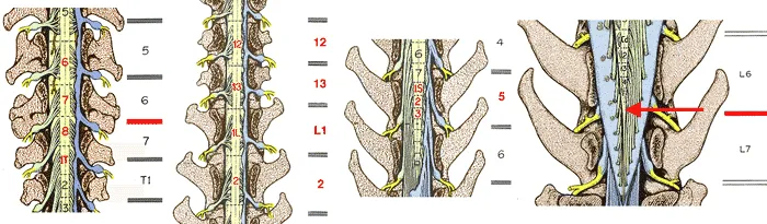
Spinal Cord and Meninges Indian Medical PG Question 3: In adults, the spinal cord normally ends at what level?
- A. Lower border of L3
- B. Lower border of S1
- C. Lower border of L5
- D. Lower border of L1 (Correct Answer)
Spinal Cord and Meninges Explanation: ***Lower border of L1***
- In adults, the **spinal cord** typically terminates at the level of the **L1 vertebral body**, or specifically, its lower border [1].
- This marks the anatomical transition from the solid spinal cord to the **conus medullaris**, which then continues as the **cauda equina** [1].
*Lower border of L3*
- While the spinal cord in **newborns** can extend as low as L3, it retracts with growth, and this level is incorrect for adults.
- An adult spinal cord ending at L3 would be considered an **abnormal finding**, potentially indicating a **tethered cord syndrome**.
*Lower border of S1*
- The spinal cord never extends to the S1 level in healthy individuals, even in newborns.
- The **sacrum (S1-S5)** is well below the normal termination point of the spinal cord.
*Lower border of L5*
- The spinal cord typically terminates well above L5 in adults.
- The **cauda equina**, not the spinal cord itself, extends through the lumbar and sacral regions to L5 and beyond.
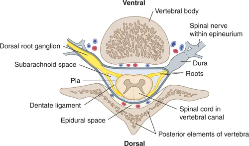
Spinal Cord and Meninges Indian Medical PG Question 4: In spinal anesthesia, the needle is pierced up to which space?
- A. Subarachnoid space (Correct Answer)
- B. Intrathecal space
- C. Epidural space
- D. Subdural space
Spinal Cord and Meninges Explanation: ***Subarachnoid space***
- In **spinal anesthesia**, the anesthetic agent is injected directly into the **cerebrospinal fluid (CSF)**, which is located in the subarachnoid space.
- This space is targeted to achieve rapid and widespread blockade of spinal nerves, leading to anesthesia and paralysis below the level of injection.
*Epidural space*
- The **epidural space** is located outside the **dura mater** and contains fat and blood vessels; it is targeted in **epidural anesthesia**, not spinal anesthesia.
- Anesthetic agents in the epidural space provide a slower onset and a more segmental block compared to spinal anesthesia.
*Intrathecal space*
- The term **intrathecal space** broadly refers to the space containing CSF, which includes the subarachnoid space, but is a less precise anatomical term for the site of injection in spinal anesthesia.
- While technically correct in referring to an injection into the CSF, "subarachnoid space" is the specific anatomical term for where the needle tip rests.
*Subdural space*
- The **subdural space** is a potential space between the **dura mater** and the **arachnoid mater**; it is not the intended target for either spinal or epidural anesthesia.
- Accidental injection into the subdural space during spinal or epidural procedures can lead to an unpredictable block with delayed onset and variable spread.
Spinal Cord and Meninges Indian Medical PG Question 5: Sensations of pain from teeth and temperature are carried by
- A. Lateral spinothalamic tract (Correct Answer)
- B. Trigeminal nerve pathway
- C. Ventral spinothalamic tract
- D. Corticospinal tract
Spinal Cord and Meninges Explanation: ***Lateral spinothalamic tract***
- The **lateral spinothalamic tract** primarily carries sensations of **pain and temperature** from the body to the brain.
- This pathway is crucial for transmitting these somatosensory modalities from the periphery, including dental structures, up the spinal cord to the **thalamus** and then to the cerebral cortex.
*Trigeminal nerve pathway*
- The **trigeminal nerve (CN V)** is responsible for sensory innervation of the face, including teeth, and jaw motor function.
- While it transmits sensory information from the teeth, its central pathway eventually synapses with the **trigeminal lemniscus** which then projects to the thalamus, rather than directly being the spinothalamic tract itself.
*Ventral spinothalamic tract*
- The **ventral (anterior) spinothalamic tract** primarily carries sensations of **crude touch and pressure**.
- It does not significantly contribute to the transmission of pain and temperature, which are the main sensations from teeth and temperature described.
*Corticospinal tract*
- The **corticospinal tract** is a major **motor pathway** that originates in the cerebral cortex and descends to the spinal cord.
- It is responsible for **voluntary fine motor control** of the limbs and body, having no role in carrying sensory information like pain or temperature.
Spinal Cord and Meninges Indian Medical PG Question 6: Ophthalmic artery is a branch of which part of the internal carotid artery?
- A. Cavernous
- B. Cervical
- C. Petrous
- D. Cerebral (Correct Answer)
Spinal Cord and Meninges Explanation: ***Cerebral (Supraclinoid)***
- The **ophthalmic artery** is the first major branch of the **cerebral (supraclinoid/C6) segment** of the internal carotid artery.
- It arises **immediately after** the ICA pierces the dura mater and exits the cavernous sinus, entering the **subarachnoid space**.
- The ophthalmic artery enters the orbit through the **optic canal** alongside the optic nerve, supplying the eye and orbital structures.
- This is the **most clinically important branch** arising from this segment before the terminal bifurcation into anterior and middle cerebral arteries.
*Cavernous*
- The **cavernous segment (C4)** courses through the cavernous sinus and gives rise to small branches like the **meningohypophyseal trunk** and **inferolateral trunk**.
- These branches supply the pituitary gland, cranial nerves, and dura mater.
- The ophthalmic artery does **NOT** arise from this segment; it arises after the ICA exits the cavernous sinus.
*Cervical*
- The **cervical segment (C1)** extends from the common carotid bifurcation to the entrance of the carotid canal at the skull base.
- This segment typically has **no branches**, serving primarily as a conduit.
- The ophthalmic artery arises much more superiorly, intracranially.
*Petrous*
- The **petrous segment (C2)** lies within the petrous part of the temporal bone in the carotid canal.
- It gives rise to small branches like the **caroticotympanic** and **vidian arteries** that supply the middle ear and pterygoid canal.
- The ophthalmic artery is not a branch of this segment.
Spinal Cord and Meninges Indian Medical PG Question 7: All are pierced in Lumbar Puncture except:
- A. Interspinous Ligament
- B. Ligamentum Flavum
- C. Supraspinous ligament
- D. Posterior longitudinal ligament (Correct Answer)
Spinal Cord and Meninges Explanation: ***Posterior longitudinal ligament***
- The **posterior longitudinal ligament** runs along the **posterior surface of the vertebral bodies**, forming the **anterior wall of the spinal canal**.
- A lumbar puncture needle **does not reach this ligament** as it enters from the **posterior aspect** of the spinal canal.
*Interspinous Ligament*
- The **interspinous ligament** is located between the **spinous processes of adjacent vertebrae**.
- It is **pierced** during a lumbar puncture as the needle advances through the posterior elements to reach the spinal canal.
*Ligamentum Flavum*
- The **ligamentum flavum** connects the **laminae of adjacent vertebrae**.
- This ligament is **pierced** by the needle just before it enters the epidural space and then the subarachnoid space during a lumbar puncture.
*Supraspinous ligament*
- The **supraspinous ligament** runs along the tips of the **spinous processes**.
- It is the **first ligament pierced** by the needle as it enters the skin and subcutaneous tissue during a lumbar puncture.
Spinal Cord and Meninges Indian Medical PG Question 8: Fluid-filled cavity at the center of the spinal cord is seen in:
- A. Subacute combined degeneration (SACD) of cord
- B. Brown Sequard syndrome
- C. Tabes dorsalis
- D. Syringomyelia (Correct Answer)
Spinal Cord and Meninges Explanation: ***Syringomyelia***
- **Syringomyelia** is characterized by the formation of a **syrinx**, a fluid-filled cavity or cyst, within the spinal cord, most commonly in the cervical region
- This cavity expands over time, compressing and damaging nerve fibers from the inside out, leading to progressive neurological deficits
- Classic presentation includes **dissociated sensory loss** (loss of pain and temperature sensation with preserved touch and proprioception) in a "cape-like" distribution
*Subacute combined degeneration (SACD) of cord*
- SACD is primarily caused by **Vitamin B12 deficiency** and involves demyelination of the **dorsal and lateral columns** of the spinal cord [1]
- Does not present with a fluid-filled cavity but rather with neuronal degeneration and demyelination [1]
- Clinical features include both sensory ataxia (dorsal column) and spastic paraparesis (lateral corticospinal tract) [2]
*Brown Sequard syndrome*
- Results from **hemitransection (damage to one side)** of the spinal cord, typically from trauma or mass lesions
- Leads to ipsilateral motor paralysis and loss of proprioception, with contralateral loss of pain and temperature sensation
- Involves damage to specific tracts rather than formation of a central cavity
*Tabes dorsalis*
- **Tabes dorsalis** is a late manifestation of **tertiary syphilis**, causing degeneration of the **dorsal columns** and dorsal nerve roots
- Characterized by sensory ataxia, lightning pains, Argyll Robertson pupils, and Charcot joints
- Does not involve a fluid-filled cavity but rather progressive demyelination
**References:**
[1] Cross SS. Underwood's Pathology: A Clinical Approach. 6th ed. Common Clinical Manifestations Of Central And Peripheral Nervous System Disease, pp. 716-717.
[2] Kumar V, Abbas AK, et al.. Robbins and Cotran Pathologic Basis of Disease. 9th ed. The Central Nervous System, pp. 1306-1307.
Spinal Cord and Meninges Indian Medical PG Question 9: Common deformity in Chiari II malformation is -
- A. Syringomyelia (Correct Answer)
- B. Hydrocephalus
- C. Meningo myelocele
- D. All of the options
Spinal Cord and Meninges Explanation: ***Syringomyelia***
- **Syringomyelia** is a common deformity associated with Chiari II malformation, characterized by a **fluid-filled cyst (syrinx)** within the spinal cord.
- This cyst can expand and damage the spinal cord, leading to symptoms such as **pain**, **weakness**, and **sensory deficits**.
*Hydrocephalus*
- While **hydrocephalus** (excess CSF in the brain) is frequently seen with Chiari II malformation, it is a **complication** or associated condition rather than a specific deformity caused by the malformation itself [1].
- It often results from the **obstruction of CSF flow** due to the displacement of hindbrain structures [1].
*Meningo myelocele*
- **Meningomyelocele** is a severe form of **spina bifida** where the spinal cord and its coverings protrude through an opening in the spine.
- It is often associated with Chiari II malformation, as they share a common developmental origin, but it is a primary **neural tube defect**, not a deformity specifically *caused by* Chiari II.
*All of the options*
- While all three conditions (syringomyelia, hydrocephalus, and meningomyelocele) are often seen in conjunction with Chiari II malformation, only **syringomyelia** is directly considered a "deformity" or direct consequence resulting from the herniation of brain tissue characteristic of Chiari II.
- Hydrocephalus and meningomyelocele are either associated conditions or complications, rather than a direct structural deformity of the brainstem and cerebellum.
Spinal Cord and Meninges Indian Medical PG Question 10: A young child with recurrent bacterial meningitis should be clinically evaluated for the presence of
- A. Spina bifida occulta with a dermal sinus tract (Correct Answer)
- B. Hypoplastic left heart syndrome
- C. Syringomyelia of the lower cervical cord
- D. Holoprosencephaly
Spinal Cord and Meninges Explanation: ***Spina bifida occulta with a dermal sinus tract***
- A **dermal sinus tract** provides a direct pathway for skin flora to access deeper structures, including the **meninges**, leading to recurrent bacterial meningitis
- This condition arises from incomplete closure of the neural tube and is often associated with cutaneous stigmata such as a **dimple, tuft of hair, or hemangioma** in the lumbosacral region
- Clinical examination should focus on the **midline back** for these telltale signs
- This is the **most common anatomical cause** of recurrent bacterial meningitis in children
*Hypoplastic left heart syndrome*
- This is a congenital heart defect resulting in an underdeveloped left side of the heart, leading to cyanosis and heart failure
- It is not directly associated with an increased risk of recurrent bacterial meningitis
- This is a **cardiac anomaly**, not a CNS communication defect
*Syringomyelia of the lower cervical cord*
- Syringomyelia involves the formation of a fluid-filled cavity (syrinx) within the spinal cord, typically causing neurological deficits related to pain, temperature sensation, and motor weakness
- While it is a neurological condition, it does **not disrupt the meningeal barrier** and therefore does not explain recurrent bacterial meningitis
- This is an **intramedullary lesion** without external communication
*Holoprosencephaly*
- This is a severe condition where the forebrain fails to develop into two hemispheres, leading to various craniofacial anomalies and significant neurological impairment
- It is a developmental brain abnormality and is **not a cause** of recurrent bacterial meningitis
- While severe, it does not create a pathway for bacterial entry into the CNS
More Spinal Cord and Meninges Indian Medical PG questions available in the OnCourse app. Practice MCQs, flashcards, and get detailed explanations.


 oka
oka