
Abdominal Trauma Imaging — Flashcards

What is the imaging of choice for diagnosing appendicitis in adults?_____
_____ sign is seen in CT because in acute pancreatitis perinephric fat is not involved
What is the best investigation for pneumoperitoneum?
Christmas tree bladder or pine cone bladder is a cystogram appearance which usually seen in severe _____ with increased sphincter tone.
_____ is the investigation of choice in Acute pancreatitis
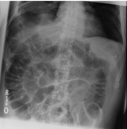
Northern exposure sign represents dilated sigmoid colon that extends cranial to the transverse colon, seen in _____ on a supine abdominal x-ray
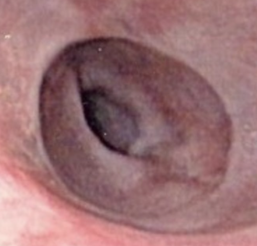
The given image shows the presence of a _____ in lower end of esophagus.
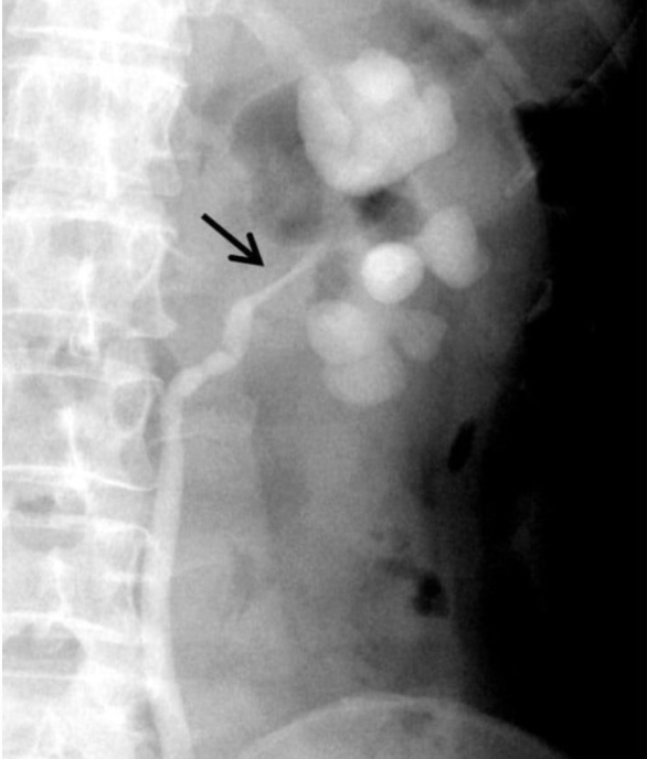
The given Retrograde ureterogram showing dilated clubbed calyces with stricture of the left ureteropelvic junction is suggestive of _____
In _____ sign or double wall sign, inner mucosal and outer serosal layers of bowel are enhanced in pneumoperitoneum
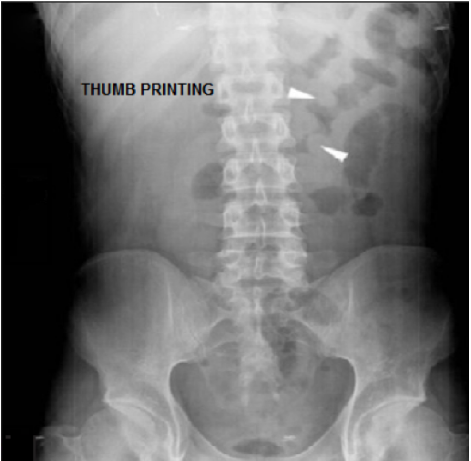
Thumbprinting sign is suggestive of _____
Abdominal Trauma Imaging Flashcards for NEET-PG
Study 10 flashcards on Abdominal Trauma Imaging for NEET-PG Radiology. These active recall cards cover the key concepts, clinical associations, and high-yield facts from this chapter of Abdominal and Pelvic Radiology. Each card is designed to test your understanding rather than just recognition, building stronger and more durable memories for exam day.
For personalised spaced repetition scheduling and unlimited flashcards, download the Oncourse app.
Frequently Asked Questions
Are Abdominal Trauma Imaging flashcards free?
How many flashcards does this chapter have?
How should I use these flashcards for NEET-PG?
Are there more flashcards for Abdominal and Pelvic Radiology?
Want unlimited flashcards?
Get full access to all flashcards, spaced repetition, and progress tracking.
Start For Free